Organelles: Components of the Human Cell

The Human Cell: Cell Illustration If you shrunk your body three million times you would be surrounded by 30


Today is my 20th anniversary as Laura Maaske – Medimagery LLC. I am grateful to a career and clients who have allowed me to be curious every day, and to render that curiosity in a way (I hope) to make complicated ideas just a little clearer and easier to understand. I celebrate this special day by talking about what has somehow been among the top ten objects I love to draw: the human cell. While cells are usually deeply networked and in connection with those around them. They are also independent units and the smallest unit of the story of what identifies an organism. Every day in my work I have the chance to learn new things, to explore, and to reveal information in a new, beautiful way. So here it goes, components of the human cell, and some drawings of organelles…


The Human Cell: Cell Illustration

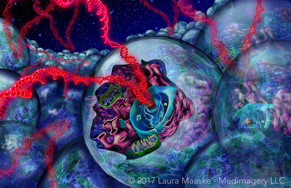
If you shrunk your body three million times you would be surrounded by 30 trillion cells made up of DNA from your own body. But as most of these cells are red blood cells, there are only 5 trillion tissue cells. You would soon notice, via the electrical surges, a profoundly busy network in communication around you.


The DNA serves as the most intimate marker of your personal identity; a reproducible basic signature of who you are.


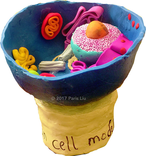
A typical animal cell, sliced open to reveal cross-sections of organelles. 
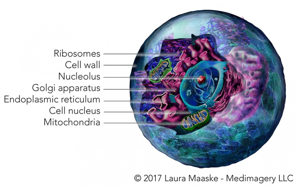


Cell organelles


What are Organelles?


Biologists liken the substructure of a cell to the organs of the body. Hence the name organelle. Inside a cell are many smaller, specialized parts, each with a specialized function for the cell as a whole. These structures are called organelles.


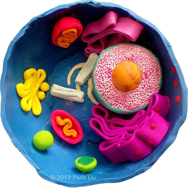
Contents of the Cell: Organelles


The Cell Wall or Cell Membrane is a two layer bilipid layer made up of proteins and carbohydrates. It sserves to offer a protective wall, blocking passage of materials in and out in a selective way. The membrane can do this because it is semi-permeable. It is specifically evolved to allow the passage of specific materials; this is achieved through both active and passive processes. The Cell Wall has a marvelous capacity to break open and closed, to fuse and to rearrange itself like a three dimensional linking necklace.


double layered cell membrane

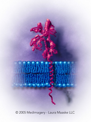
A bilipid cell membrane structure.


The Nucleolus is not technically an organelle because it lacks a menbrane. This is a specialized area where DNA is held and messages emerge.


Ribosomes are small particles, drawn above as yellow dots, which float in the Endoplasmic Reticulum and also are attached along the membrane of the endoplasmic reticulum. Ribosomes functiona as the energy factories of the cell. They produce RNA proteins.


Golgi Apparatus or Golgi Body has extensive membrane surface that is highly reformable. Its bilipid membrane breaks or pinches off into smaller or larger vesicles. The golgi apparatus stores proteins and carries them to the cell wall to be released. Under the microscope, the golgi apparatus has a stacked appearance like pancakes.


The Nucleus is the largest component of the cell. Like the Cell Wall, it is surrounded by a bilipid membrane. And like the Cell Wall, it is semi-permeable by active and passive means. Like the cell wall, the nucleus has a wonderful capacity to rearrange itself, to enlarge, to shrink, or to reunite with its membrane envelope components. At the heart of the nucleus is the great treasure of the cell, the DNA. As such, its function is to regulate the duplication and reproduction of the cell itself.


Mitochondria are another folded structure, but the folds are contained within a oval or round vesicle. The mitochondria are responsible for energy metabolism. This organelle plays a role in intracellular digestion. Cells that use more energy, such as muscle cells, have more mitochondria to offer a greater facility for energy consumption.
 
 
Mitochondria cross-section

Mitochondria cross-section


Cell Tissue Types


The specialized organelles within the cell perform varied basic functions to carry out the needs of the cell, proving providing for the specialized needs of each tissue. In fact there are over 200 different cell types in the body, including 4 tissue types:


  1. Epithelial Cells for protecting surfaces and lining hollow structures, skin, veins, arteries, edges of organs. They can also form glands. These cells initiate their own death as they rise to the surface, in order to create a protective layer from the outside world.
  2. Muscle Cells specialized in helping the organism move. It can contract and adjust the shape and diameter of the body at large.
  3. Nervous tissue cells for transmitting messages by electrical signaling. These are neurons or accesory cells to neurons.
neuron illustration

Nerve cell with its long axon to conducting messages. Ridges show the fatty myelin cells which coat the axon sheath to allow faster transport and greater conductivity.

  1. Connective tissue, dense with proteins for creating structure to hold the organism together. Connective tissue provides immune functions. This cell type includes red blood cells.


Like atoms to the molecules, cells are the building blocks of living organisms. I didn’t create an animation for the cell above, which makes it seem serenely quiet and still, when it is full of active life and constant movement. Every minute more than 200 million cells in our body die, and another 200 born. Of course, this is a rough estimate and the number declines with age.


New Findings in Cell Anatomy


Cell biology was mostly discovered in the 1940s. But there was recently a new discovery. In June 2015, scientists discovered a never known structure they call the “mesh”. The mesh is a structure made up of microtubules, that holds cells together.


Learning About the Cell

 
My daughter is a high school freshman. For biology class she was asked to create a cell structure. I thought I’d include her model in this article for anyone who is visiting this site to learn about the cell:


_________________________________

September 20, 2017

Laura Maaske, MSc.BMC.

Biomedical Communicator

Medical Illustrator

Medical Animator

Health App Designer


Medimagery Medical Illustration

___________________________________
Laura Maaske – Medimagery LLC
Medical Illustration & Design
___________________________________


Bye bye!!!

cell window components

[jp_post_view]


Pin It

About Laura

Medical Illustrator; science and art lover.

2 thoughts on “Organelles: Components of the Human Cell

    1. AUTHOR

Comments are closed.