Illustrated Gastric Stomach Layers: The Three Layers of the Stomach

3 smooth muscle layers of the stomach, together are called the muscularis externa. They are made up of


Stomach Layers: the Gastric Layers

Illustrated Gastric Stomach Layers: The Three Layers of the Stomach


First: a Little Basic Anatomy

What is the Stomach?


The stomach is a hollow, muscular organ on the upper left side of the abdomen, with one entrance and one exit. One leads in from the esophagus. And the other leads out into the duodenum. The door in is surrounded by the edge of the diaphragm. Food arrives to the stomach from the mouth after it has traveled down the esophagus. When food arrives at the end of the esophagus, it enters the stomach through a muscular valve called the lower esophageal junction (GEJ).


3 stomach layers

This gastric illustration depicts the three layers of smooth muscle which line the stomach wall.


This gastric illustration depicts the three layers of smooth muscle which line the stomach wall.


There are four main regions in the stomach: the cardiac region, the fundus, the body, the pylorus, and the duodenum. The cardiac region is where the esophagus connects to the stomach, and it is the door to the stomach. The dome-shaped fundus is the top of the stomach and lies just below the diaphragm. Under the funds, and comprising the large central portion of the stomach, is the body of the stomach, Inferior to the body is the  funnel-shaped pylorus. The pylorus connects the stomach to the duodenum. The smooth muscle pyloric sphincter is a contracted for at the bottom of the stomach, which regulates the passage of broken down food.


The interior of the stomach is a rough surface of large folds, ridges of muscle, called rugae. The stomach secretes acid and enzymes that digest food. The stomach muscles contract periodically, churning food to enhance digestion. The pyloric sphincter is a muscular valve that opens to allow food to pass from the stomach to the small intestine.


Being made of muscle, the stomach is a highly active structure, contracting and continually changing position and size. In fact, the only time it appear in shape as I have drawn it here is when it is distended, or full. Rhythmic contractions create mechanical assistance to digestion. When empty, the stomach is about the size of your fist.  And it can stretch to hold as much as 4 liters of food and fluid, or more than 75 times its empty volume. Carbohydrate digestion begins in the mouth, but that is minimal. Carbohydrate digestion really occurs in the stomach. The stomach begins the digestion of proteins and fats. But very few nutrients are absorbed directly through the stomach. Some alcohol may be absorbed in the stomach.

Another important function of the stomach is to act as a waiting room or holding chamber for food. First food is taken into the stomach and mixed with digestive juices until they are converted into chyme. Then the mixture is slowly released to the small intestine. The stomach can receive huge quantities of food at a time. But the small intestine cannot. The small intestine can only function by taking in food slowly, bit by bit, from the stomach. 


Why does the stomach have 3 layers of muscle?

Smooth muscle in the stomach contracts during digestion, in order to break down nutrients. The muscle layers in the human stomach are each striated in different directions. Because smooth muscle can only contract in one direction, it is functionally adaptive for the stomach, which must contract as an organ overall, to contract in three directions. Another muscle in the body which has muscle fibers running in different directions is the tongue. The stomach is the only organ in the digestive system to have three muscle layers. The rest of the gastrointestinal tract, or GI tract, contains only two muscle layers.
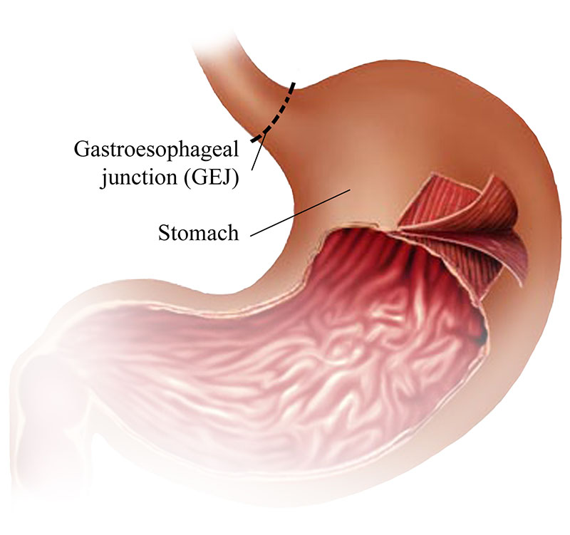


The Muscular Layers of the Stomach and Muscularis Externa

The stomach wall is comprised of the same four layers as most of the rest of the alimentary canal, but with some differences in the mucosa. In addition to the circular and longitudinal smooth muscle layers which are found throughout the digestive system, the muscularis includes an inner oblique smooth muscle layer. As a result, the stomach muscle is capable to churn food, mechanically breaking it down into a slurry, in addition to moving food through the canal. The muscles together as a group, are called the muscularis externa


  1. Muscle layer 1: outer longitudinal layer – This layer is composed of smooth muscle, continuous with the smooth muscle which surrounds the esophagus. Below this longitudinal muscle is the Auerbach’s plexus, or myenteric plexus, above the middle circular. It is a layer of nerves which cause peristalsis, movement of the muscle itself. The peristalsis allows food to mix. 


  1. Muscle layer 2 – middle circular layer – This layer of smooth muscle is called the circular layer and lies beheath the longitudinal muscle layer. It wraps around the body of the stomach and extends downwards to form the pyloric sphincter. It wraps in a circular orientation around the pylorus, and is held in a constricted state normally. The normal constriction of this muscle is what creates the pyloric sphincter, which controls the movement of chyme into the duodenum. This layer is concentric to the longitudinal axis of the stomach. (note: chyme is a thick and acidic mixture of acidic digestive fluids and partially digested food, which moves from the stomach to the small intestine as digestion moves along the digestive tract.)


  1. Muscle layer 3 – inner oblique layer – This layer of smooth muscle is the middle layer called the circular layer. It wraps around the body of the stomach, extending downward to form the pyloric sphincter along with the circular muscle layer. This layer is responsible for creating the motion that churns and physically breaks down the food. It is the only layer of the three which is not seen in other parts of the digestive system. The antrum has thicker skin cells in its walls and performs more forceful contractions than the fundus.


Microscopic View


Figure 2. The stomach wall is adapted for the functions of the stomach. In the epithelium, gastric pits lead to gastric glands that secrete gastric juice. The gastric glands (one gland is shown enlarged on the right) contain different types of cells that secrete a variety of enzymes, including hydrochloride acid, which activates the protein-digesting enzyme pepsin.

The stomach mucosa’s epithelial lining consists only of surface mucus cells, which secrete a protective coat of alkaline mucus. A vast number of gastric pits dot the surface of the epithelium, giving it the appearance of a well-used pincushion, and mark the entry to each gastric gland, which secretes a complex digestive fluid referred to as gastric juice.

Although the walls of the gastric pits are made up primarily of mucus cells, the gastric glands are made up of different types of cells. The glands of the cardia and pylorus are composed primarily of mucus-secreting cells. Cells that make up the pyloric antrum secrete mucus and a number of hormones, including the majority of the stimulatory hormone, gastrin. The much larger glands of the fundus and body of the stomach, the site of most chemical digestion, produce most of the gastric secretions. These glands are made up of a variety of secretory cells. These include parietal cells, chief cells, mucous neck cells, and enteroendocrine cells.


To read more about the stomach and other organs of the abdomen and to see more illustrations, read my article, “Medical Illustrations of Abdominal Organs & Digestive System


Stomach Ailments: Diaphragmatic Hernia
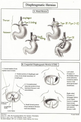


THe liver lies in front of the stomach. A small portion of the stomach is visible here beneath the liver and above the transverse colon


May 16, 2017

Laura Maaske, MSc.BMC. Biomedical Communicator


Medimagery Medical Illustration

___________________________________
Laura Maaske – Medimagery LLC
Medical Illustration & Design
___________________________________

Text Copyright © 2017 Medimagery – Laura Maaske LLC

Illustration © 2000 Columbia Healthcare EHC, illustrated by Laura Maaske – Medimagery LLC.


Pin It

About Laura

Medical Illustrator; science and art lover.