
Visions of the Prenatal Development: Human Embryo & Fetal Illustrations
Embryo Illustration and Fetus Illustration Series
Unfolding of New Life | Embryo, Fetus, & Newborn Baby
Writing and Fetus Illustrations drawn by Laura Maaske, MSc.BMC, Medical Illustrator & Medical Animator
What follows is a series of illustrations and animations I prepared for Lamaze to depict the development of the human embryo and fetus. The series offered a range from Week 6 to Week 40. But I wanted my writeup to be more complete. So I have added earlier development of the embryo as well as the gametes: sperm and egg cells.
Even in our modern technologically driven age, the world in which the embryo develops and the fetus lives is still a place which inspires our imaginations and our wonder at the rapidity and vast changes taking place. These illustrations, which I prepared individually for Lamaze, offered me a chance to explore those questions in greater depth by creating a full set of embryo illustrations and fetus illustrations.
Click to Play Video
The illustrations in the video above reveal fetal development beginning at 6 weeks of gestational age and progressing through each week, with summary text, until the final 40th week of pregnancy. At each week vital information about development is offered, along with a comparatively sized vegetable or fruit, to mark the size of the growing baby.
Basic Development: Egg and Sperm Illustrations
Egg and sperm are initially produced through a process called meiosis. Sperm are produced in the male testes. Cells with 46 chromosomes, called diploid cells, divide in such a way as to produce cells with 23 chromosomes, called haploid cells. The result is haploid sperm cells containing 23 chromosomes.

In females, diploid cells, with 46 chromosomes, in the ovary, divide to produce haploid cells. These haploid cells are eggs, each of which contains 23 chromosomes.
Sperm migrate from the testes through and out the penis during sex. Eggs, one of which is produced each month, migrate from the ovary to the uterus once a month. Egg and sperm typically meet in the fallopian tube, and migrate to the uterus over a course of 5 days. Eggs which do not meet sperm are discharged at the time of a woman’s menstruation.

During a single ejaculation, a human male can release from 50 million to 1 billion spermatozoa. Typically, only one of these spermatozoa will achieve entry to an egg. When an egg cell becomes fertilized with a sperm cell, the two haploid cells unite and integrate their chromosomic material to become a diploid cell, a fertilized egg. The gender of the future baby is determined by the male genetic contribution, at the time of conception. The diploid cell is the first cell of a human body. It holds 46 chromosomes, half contributed by each of its parent cells. This 46 chromosome cell then divides by the mitotic process, mitosis, thus producing a two celled organism. Each of these calls divides and grows, thereby beginning the human life as a multicellular organism.

To learn more about the development of gametes, view my illustrated article here.
First Trimester Development with Embryo & Fetus Illustrations
The Embryonic Stage
Early Embryogenesis: Growth into an Embryo
As these first 46-chromosome cells multiply, they create a zygote. Through this process of mitosis, the cell multiplies to produce an embryo: a new and complete human being. It will be called an human embryo once it has implanted in the uterus; into the uterine endometrial lining. Even this is not guaranteed, as only 60% of embryos implant.
This prenatal series for Lamaze focused on the earliest stages of development, including the processes of early gene expression, histogenesis, cleavage, blastulation, gastrulation, neurulation and organogenesis: the most rapid period of growth. And in fact, I’ve never been requested to illustrate these stages. New parents don’t express as much curiosity about these stages, it seems, because they have passed by the time a woman is aware she is pregnant. I certainly hope there are some new research developments in this area, to propel a client request. It is my favorite stage of development. The changes are remarkable and life at this stage looks a bit like something we might imagine on an alien landscape.
My references to developmental time here are classified as embryonic age, which is the age from fertilization. Gestational age is the age from the mother’s last menstruation, and this would make the embryo seem up to 2 weeks older, as it can take 2 weeks for fertilization to occur.
6 Week Old Embryo Development and Embryo Illustration
At 6 weeks an embryo is quite curled up, like the letter “C”. This is a rapid period of prenatal development, with a phenomena to-do list. Note the pharyngeal arches, in the embryo below. These are a series of ridges around the “neck” and “chin” region. They are a vestige of earlier stages in evolution. In fish, for example, these pharyngeal arches develop into gills. In humans, the pharyngeal arches develop into anatomical structures of the mouth, neck, face, and ear. At six weeks of age, an embryonic is newly revealing tiny lung bud, and its heart begins to beat in a regular rhythm. The spleen is new. The embryo is just beginning to exhibit arm buds and ear pits. And the rectal and urinary tubes begin to separate. A yolk sac is in place and provides nutrients during the embryonic period before nutrition first arrives from a placenta. The embryo measures about 4 mm (1/8 inch) from head to rump, and is about the size of a grain of rice.

A developing baby is considered to be an embryo between the Fifth and Eleventh Week of development.
7 Week Embryo Development and Embryo Illustration
At 7 Weeks the embryo is 10,000 times bigger at this time than it was at the moment of conception. Buds form on the embryo’s developing arms which look like hand paddles. These will become the hands. Legs and a new stomach are are developing, too. Blood is first beginning to circulate in the tiny life. The kidneys are built and show the first signs of filtering toxins. The eyes and nose are beginning to form as optic cups and nasal pits. The brain forms into five separate vesicles. The embryo measures 8mm (0,315 inch), and is about the size of an cucumber seed.*
8 Week Embryo Development and Embryo Illustration
At 8 Weeks the lungs are beginning to form, as the brain further enlarges. Arms and legs grow longer with the beginning signs of fingers and toes. The sex organs begin to develop. And the embryo is 13 mm (1/2 inch) long, and about the size of a watermelon seed.
The Fetal Period Begins
At What Age does an Embryo become a Fetus? Here’s the Answer?
9 Week Embryo Development and Embryo Illustration
At 9 Weeks the heartbeat is audible using a doppler. The embryo is beginning to make some movements of its limbs. It is possible to see now the place where elbow and toes are developing. Nipples on the chest and tiny hair follicles are visible. By this time, all the basic human organs are present and under development. The eyes are more than cups, with lids, but they are fused shut until 27 weeks. The embryo is about 18 mm (3/4 inch) long, which is the size of an almond.

10 Week Embryo Development and Embryo Illustration
The fetus at 10 weeks has a fully developed outer ear and a real chin. It has a bulging forehead. Her forehead is temporarily bulging, the outer part of her ear is fully developed and her chin is no longer bent down to her chest. Arms and legs are capable of rotation at the shoulder joint and hip joints. The heart is now almost fully formed and is beating at about three times the rate of an adult heart. Fingernails first appear. The yolk sac begins to disintegrate. The fetus measures about 30 mm (1.2 inches) in length, from head to rump, and is roughly the size of a grape.

11 Week Embryo Development and Embryo Illustration
At 11 weeks an embryo has graduated to the fetal stage, defined as the period after which all major structures are present. At this time, the digestive system, endocrine system, lymphatic system, muscular system, nervous system, urinary system, circulatory system of arteries and veins, integumentary system, reproductive system, and respiratory system with lungs are growing and developing their complex structures. The mouth is developing tiny milk teeth within the gumps, and the bones of the palate on the roof of the mouth are fusing. Fingers and toes are no longer webbed, and, while too tiny to be felt by the mother, the fetus is beginning to kick its arms and legs rapidly, and it can now open and close its hands.
The fetus is not so sensitive to chemical and general environmental changes, because the organs and anatomical structures which depend on chemical signals to initiate specific developmental triggers are already under development. However, the fetus is still vulnerable to toxic levels of exposure to certain substances, which may cause morphological and physiological deformities or less life-endangering congenital malformations. The next weeks will be marked more by simple growth than by changes in structure. The fetus is now about 4.1cm (1.6in) from crown to rump, or about the size of a strawberry.

12 Week Fetal Development and Fetus Illustration
At 12 weeks the fetal eyes are moving closer together, giving it a more “human” appearance. The fetus shows sucking motions with its mouth now. Fetal reflexes are more advanced, so the fetus will respond on the ultrasound if the belly is lightly poked. The very first bone formation begins to take place in the cartilage structure which makes up the fetal skeleton. This process of bone formation will continue through out fetal development and childhood almost into adulthood. The fetus is about 5.4cm (2.1in) from head to rump, and is about the size of an avocado pit.

13 Week Fetal Development and Fetus Illustration
At 13 weeks the first developments of the frontal cortex are taking place. The first fingerprint patterns develop, although they are still in early formation. Fetuses at this age are sucking in and swallowing amniotic fluid as a way to practice breathing and even to absorb some nutrients: sugars, proteins, and salts, and flavors from the mother’s foods. They might be seen sucking their thumbs for the first time, on ultrasound. Any fluids absorbed are beginning to be processed by the kidneys and ureters, and even peed out back into the amniotic fluid.
A male fetus at this age has testicles and a growing penis. A female fetus at this age has ovaries with about two million eggs already. The head is still quite large and makes up a third of the length of the body. But the fetus has grown to about 7.4cm (2.9in) from head to rump, and is about the size of a lime.

14 Week Fetal Development and Fetus Illustration
At 14 weeks the fetus is continuing to practice arm and leg movements movements, which are now more in sync with one another. Facial movements first begin showing up on ultrasounds at this age. Eyes begin to move under fused eyelids. Lanugo, fetal hair, is beginning to form all over the body as a way to regulate temperature. It will disappear around 36 weeks.
The fetus is growing in such a way now that the arms and legs are catching up to the head more, reducing the proportion of the head from a third to a quarter the length of the body. The fetus measures about 8.7 cm (3.4 in) from crown to rump, and is about the size of a lemon.

15 Week Fetal Development and Fetus Illustration
At 15 weeks the fetal eyes, while still fused shut, begin to respond to light changes, and the ears are beginning to function and take in sounds in the mother’s body and from the environment outside. A 15 week old fetus is about 10.1 cm (4 in) long from head to rump, and is about the size of an apple.

Second Trimester Begins
16 Week Fetal Development and Fetus Illustration
At 16 weeks the fetus is developing muscles so that some of them can move individually. The circulatory system is fully developed and is pumping blood to all regions of the fetal body. Muscles in the neck and back allow some flexion and extension so the fetus is not always entirely curled up. The fetus may be seen on the ultrasound to grasp its hands together. The fetus and its placenta enters a tremendous growth spurt at this time, doubling its weight in the next 3 weeks. The philtrum forms above the fetus’ upper lip, to form the small indentation common to the human face. The scalp is revealing early signs of where the hair will grow in later. The fetus at this age measures around 11.6cm (4.6in) from head to rump, and is about the size of an avocado.

17 Week Fetal Development and Fetus Illustration
At 17 weeks the fetus is becoming more responsive to auditory and visual stimulation. It’s developing the fatty myelin sheath around its spinal cord and nervous system as a way to spied up the conduction of nerve signals. The skeleton is continuing to calcify from cartilage, and the limbs are growing so the proportions are looking more normal. A fetus at 17 weeks of gestational age is 13cm (5.1in) from head to rump, and is about the size of an ordinary baking potato.

18 Week Fetal Development and Fetus Illustration
At 18 weeks a baby’s gender can be seen clearly on its ultrasound. A female fetus’ fallopian tubes, ovaries, womb, and vagina are all fully developed and in their proper position. A male fetus’ penis will be clearly distinct, and its testicles will have begun to descend from their position inside the pelvis to the area of the scrotum. Ears are also in place as they will be when born. Eyebrows have formed. And the eye muscles are fully functional to turn the eyes different directions. The fetus is very responsive to sensory stimulation. A fetus at about this age is 14.2 cm (5.6 in) from head to rump, the size of a bell pepper.

19 Week Fetal Development and Fetus Illustration
At 19 weeks the fetal movements can be felt by many mothers. Inside the fetus, the areas of sensation are developing in the fetal brain: hearing, touch, vision, scent, and taste. Hair is beginning to grow on the scalp. Arms and legs are getting to their newborn proportions. A fetus at this age measures approximately 15.3 cm (6 in) from crown to rump, and is about the size of an orange.

20 Week Fetal Development and Fetus Illustration
At 20 weeks the vernix caseosa begins to coat the fetal body. This is a white substance, shiny and full of fat, which is like lotion to coat the fetus, protect it from dehydration exposed to amniotic fluid, and to help make the passage more slippery through the birth canal. A fetus at this age is measured from head to foot instead of head to rump. The size is about 25.6 cm (10 in) from head to toe, which is about the length of a banana.

21 Week Fetal Development and Fetus Illustration
At 21 weeks there’s still plenty for room for a growing fetus to move around and flex in its uterus. It sleeps as much as a newborn. It is becoming acclimated to the different tastes of the amniotic fluid, from day to day, so that it will prefer those tastes after birth. Its lips are becoming more defined. Skin is developing surface blood vessels, making the fetus appear more pink. Nerve connections forming between the brain and distal muscles are becoming more consolidated. So the fetal movements or “quickening”, are less jerky and more coordinated and controlled.
Eyebrows thicken and eyelashes develop for the first time. There are fingernails. For the first time this week, the fetus weighs more than its placenta. The fetal growth will continue to outpace its placental growth throughout the rest of the pregnancy. Lanugo covers the entire fetal body by now. At this time the fetal heartbeat is loud enough to be detected with a stethoscope. A fetus at 21 weeks is about 26.7 cm (10.5 in) long and 11.5 oz in weight, and is about the size of an ordinary sweet potato.

22 Week Fetal Development and Fetus Illustration
At 22 weeks the nutrients absorbed from the amniotic fluid and placental circulatory system are beginning to enter the fetal digestive system in small amounts. This will create a sticky sort of fecal material in the bowels, called meconium, which will be defecated sometime after birth.
Fetal eyes are so well developed now that the iris’s are formed and yet hold no pigment. So there is no eye color yet. Fingernails have grown rapidly so they reach the end of the fingertip now. If the fetus is a female, her mammary glands, responsible for making breastmilk, are developing.
At 22 weeks the fetus is roughly 27.8 cm (10.9in) in size, from head to toe, weighs about a pound, and is about the size of a papaya.

23 Week Fetal Development and Fetus Illustration
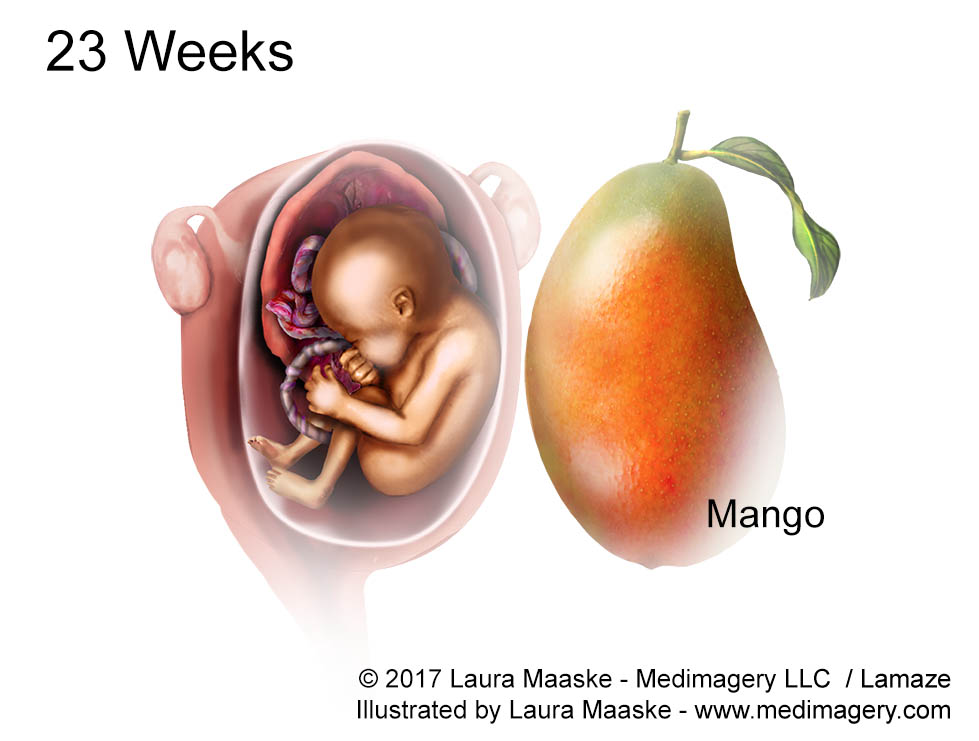
At 23 weeks the fetal heart is growing strong. Bone marrow is beginning produce new red blood cells for the first time. Up until this point, the liver and spleen were responsible for red blood cell production. Another growth spurt begins so the fetus will double its weight in the next 4 weeks.
The fetal ears are also becoming increasingly sensitive, so that sounds outside the womb such as a siren or music are becoming more clear and distinct day by day. Eyebrows and eyelashes are fully formed. The startle reflex (moro reflex) is active. And lung sacs, called alveoli, are growing in preparation for the outside world. The fetal length is about 28 cm (11.2 inches) long from head to toe. And the baby weighs about 1.1 lb. He or she is about the size of a large mango.
24 Week Fetal Development and Fetus Illustration
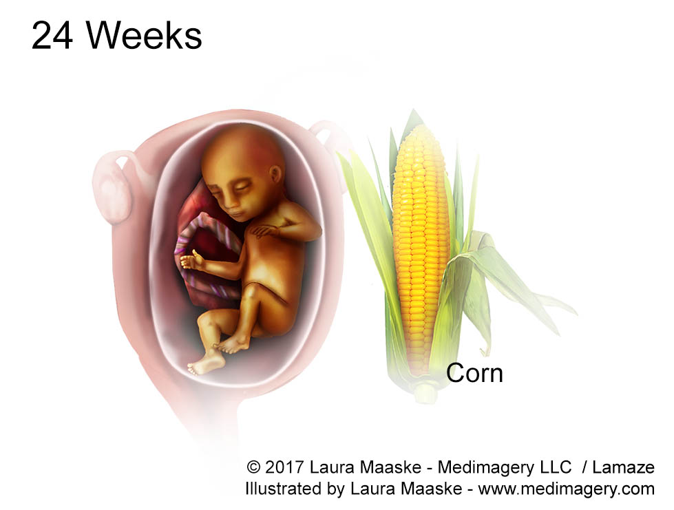
At 24 weeks the fetus is raising its eyebrows and experimenting constantly with facial expressions. His or her lungs are well enough developed now that if for some reason it needed to be born, it would have a chance to survive.
Fetal length is about 30 cm (11.8 in). Weight is about 600g (1.3lb), And the unborn child is about the size of an ear of corn.
25 Week Fetal Development and Fetus Illustration
At 25 weeks the rods and cones in the retina are beginning to develop so colors can be distinguished in bright conditions, and shadows and shapes in dark conditions. Nasal lining also delivers scent information to the brain from the amniotic fluid.
Fat is just beginning to accumulate under the skin to give protection and insulation after birth. Scalp hair is growing and the hair color is visible. Lungs are developing surfactant, a necessary substance which lines the lung and is used to aid breathing. At 25 weeks a fetus is about 34.6 cm (13.6 in) from head to toe. It weighs 660g (1.5lb) and is about the size of a cucumber.

26 Week Fetal Development and Fetus Illustration
At 26 weeks the fetus is increasingly developing blood vessel supply to the lungs, the extraordinarily complex spinal column is delaying for flexibility, and strengthening. The cochlea in the inner ear are now developed, so he or she is able to to recognize specific voices. However, the cochlea will continue to develop for speed and conductivity until the new baby is about 18 months old. The fetus is about 35.6 cm (14 in) long from head to toe, weighs about 760 g (1.7 lb), and is about the size of a rutabaga.

27 Week Fetal Development and Fetus Illustration
At 27 weeks for the first time in its life, the fetus may begin to open and close its eyes. Sleep and waking patterns show even greater regularity. And there are periods of REM sleep now, showing the same brain patterns as we experience during dreaming. The lungs are producing surfactant to make breathing efficient, so a birth at this time would pose less risk. The fetus spends more time practicing breathing. Sometimes an unborn baby at this age will respond to a spicy dish its mother has eaten, two hours later after the meal is digested, by hiccuping. Hiccups are normal and natural. A baby at 27 weeks of gestation weighs about 1.9 lb (875 g), and about 36.6 cm (14.5 in) from head to toe, which is about the size of an eggplant.

The Third Trimester
28 Week Fetal Development and Fetus Illustration
At 28 weeks the Third Trimester begins. Baby’s heart rate is slowing down to 140 beats per minute, which is still twice as fast as the adult heart. Baby’s eyes can produce tears and her or she practices blinking. Sucking capacity is greater now, as the fetus has been practicing this for some time. Even coughing is skill that the fetus practices in the womb. A fetus at 28 weeks weighs just over 1 kg (2.2 lbs). It is about 37.6 cm (14.8 in) long, and roughly the size of a butternut squash.

29 Week Fetal Development and Fetus Illustration
At 29 weeks the fetal brain, which began life as smooth, is developing folds and grooves. 200 mg of calcium are added to the fetal skeleton each day. The fetus now weighs a little over 1.1 kg (2.5 lb) and is about 38.6 cm (15.2 in) from head to toe. Baby is about the size of a head of cauliflower.

30 Week Fetal Development and Fetus Illustration
At 30 weeks baby’s eyes are developed, but only capable of focusing on very close objects. Baby weighs about 1.3 kg (2.9 lb) and measures around 40 cm (15.7 in) from head to toe, about the size of a cabbage.

Third Trimester
31 Week Fetal Development and Fetus Illustration
At 31 weeks the womb is beginning to feel snug, so the fetus will keep its legs drawn to its chest. Baby also spends more time head down, as it preps for birth. Not all babies make this turn at once. Many wait until 36 weeks. Stores of iron, calcium, and phosphorus are building up in baby’s body. The brain is developing thalamic brain connections, responsible for interpreting sensory input. Babies at this age suck their thumbs so vigorously they might be born with a callous on their thumbs. Bone marrow is now totally responsible for red blood cell production. Downy lanugo hair begins to fall off. Baby weighs about 1.5 kg (3.3 lb), and measures about 41.1 cm (16.2 in) from head to toe, and is about the size of a coconut.
32 Week Fetal Development and Fetus Illustration
At 32 weeks fingernails and toenails are fully formed. Baby will scratch himself or herself if there is an itch. Baby weighs about 1.7 kg (3.7 lb), measures roughly 42.4 cm (16.7 in) from head to toe, and is about the size of an acorn squash.


33 Week Fetal Development and Fetus Illustration
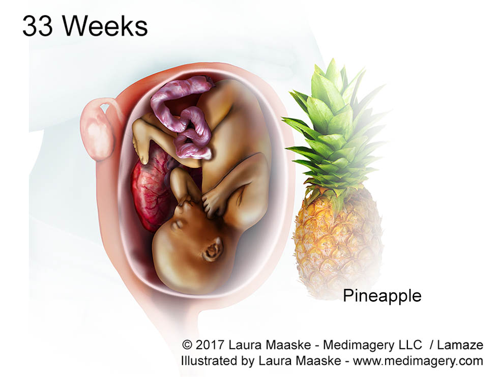
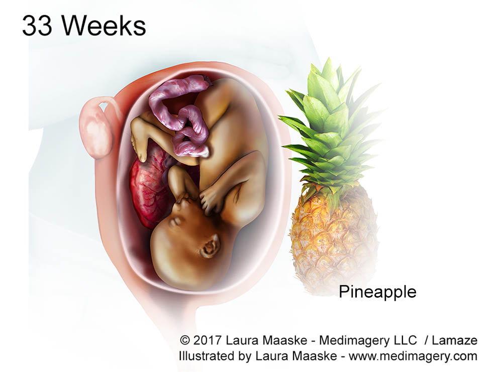
At 33 weeks the fetus is beginning to plump up with fat for insulation after birth. The uterine wall is growing stretched and thinner, allowing more light in. So baby is more sensitive to day and night. Its eyes are now closed during sleep but open during waking hours. His nervous system is developed and his immunes system is getting ready for birth. Skin is plump. Skull bone plates remain pliable to allow passage through the birth canal. Fontanelles will remain until 18 months after birth. Baby now weighs about 2 kg (4.2 lb), measures about 43.7 cm (17.2 in) from head to toe, and is about the size of a pineapple.
34 Week Fetal Development and Fetus Illustration
At 34 weeks it may be possible for baby to recognize songs and specific sounds. The amniotic fluid is at his greatest volume around now: as much as 1000 ml (1.6 pints). It will begin to decrease now so that at the time of birth the volume will be 600 ml (1 pint). A fetus at 34 weeks weighs around 2.1 kg (4.7 lb) and measures about 45cm (17.7 in) from head to toe. It is about the size of a cantaloupe.

35 Week Fetal Development and Fetus Illustration
At 35 weeks baby weighs about 2.4 kg (5.3lbs), and measures around 46.2 cm (18.2 in) from head to toe. Neurons and brin gyro and sulci continue to develop, and brain growth takes up more than half of baby’s energy.The fetus is now about the size of a honeydew melon.


36 Week Fetal Development and Fetus Illustration
At 36 weeks a birth would be considered an “early term birth”. Both lanugo and vernix caseosa are shedding. Most fetuses as this age are faced head down, in the cephalic position for birth. Some babies’ heads have dropped down into the pelvis in the engaged position, ready for birth. It gives the mother more room to breathe: called lightening. The fetus weighs about 2.6 kg (5.7 lbs) and measures around 47.4 cm (18.7 in), which is about the size of a spaghetti squash.


37 Week Fetal Development and Fetus Illustration
At 37 weeks a fetus weighs over 2.9 kg (6.3 lb), is about 48.6 cm (19.1 in) long from head to toe, and is about the size of a stalk of swiss chard.


38 Week Fetal Development and Fetus Illustration
The average weight of a baby at 38 weeks of pregnancy is roughly 3 kg (6.8 lbs), and measures about 49.8cm (19.6in). Baby is about the size of a buttercup squash.


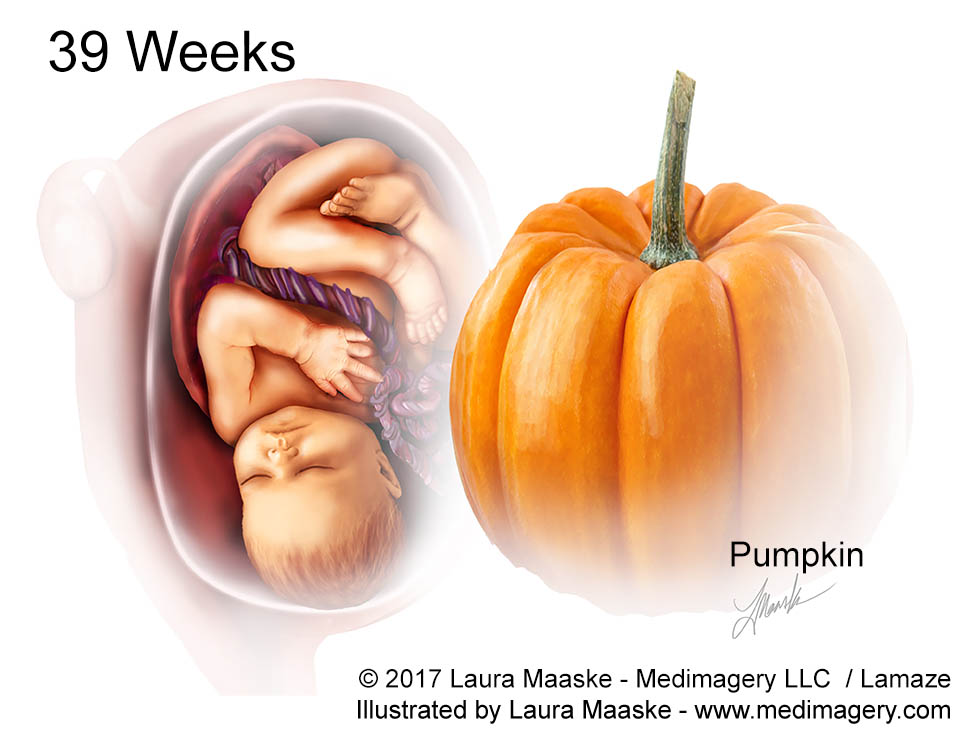
39 Week Fetal Development and Fetus Illustration
At 39 weeks. By this point, baby’s brain has billions of neurons. Baby weighs about 3 kg (6.8 lbs), and measures roughly 49.8 cm (19.6 in). Baby is about the size of a pumpkin.

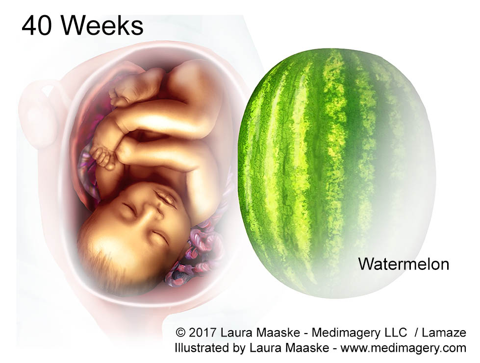
40 Week Fetal Development and Fetus Illustration
At 40 weeks baby is about 51.2cm (20.2in) from head to toe, weighs roughly 3.5 kg (7.6 lbs), and is about the size of a small watermelon.

About the Client, Lamaze
I learned many good things about Lamaze during the course of this project and with this great team. Lamaze International is a nonprofit business, endeavoring to educate in the advancement of healthy and safe pregnancies as well as early parenting. Lamaze uses evidence-based research to uncover and to advocate the best decisions, in conjunction with those advocated by the World Health Organization.
- Note that all embryological and fetal size and weight data is taken from barycenter.com
February 21, 2017
_______________________________________
Laura Maaske, MSc.BMC, Medical Illustrator & Medical Animator
e-Textbook Designer | Health Communicator | Scientific Illustrator


