
Spermatogenesis, the Development of Sperm, and the Sketch to Illustration Process
Sketching and Preparation of a Medical Illustration
Written & illustrated by Laura Maaske, MSc.BMC, Medical Illustrator & Medical Animator| e-Textbook Design
This was a particularly complicated topic for me, as a student, and here are a few of the solutions I came up with long ago, for making the topic more clear to myself and my audience:
An original sketch, spermatogenesis, created large on several sheets, taped together:
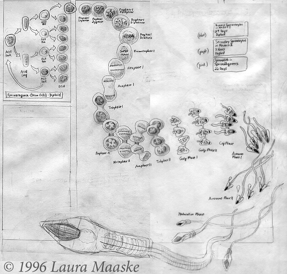
Unfortunately, this is such an old illustration, I no longer have the layout software I used to prepare the text, Aldus Pagemaker. But I think the text, however misaligned, is still helpful to understand the process of cell replication and division:
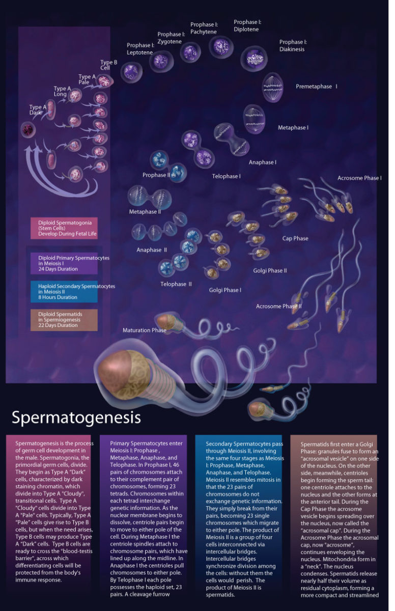
Spermatogenesis is a Meiotic Process by which Haploid Sperm Cells are Produced
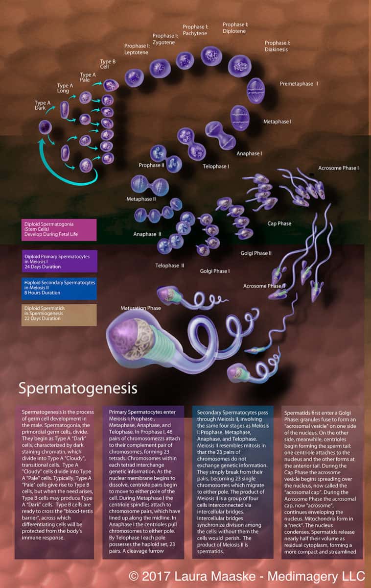

Stages of Spermatogenesis
Diploid Spermatogonia
(Sperm Cells)
Develop During Fetal Life
Early Stage 1
Spermatogenesis is the process of germ cell development in the male. Spermatogonia, the primordial germ cells, divide. They begin as Type A ?Dark? cells, characterized by dark staining chromatin. These Type A ?Dark? cells divide into Type A ?Cloudy? transitional cells. Type A ?Cloudy? cells divide into Type A ?Pale? Cells. Typically, Type A ?Pale? cells give rise to Type B cells, but when the need arises, Type B cells may produce Type A ?Dark? cells. Type B cells may produce Type A ?Dark? cells. Type B cells are ready to cross the ?blood-testis barrier?, across which differentiating cells will be protected from the body’s immune response.
Diploid Primary Spermatocytes
in Meiosis I
24 Days Duration
Stage 2
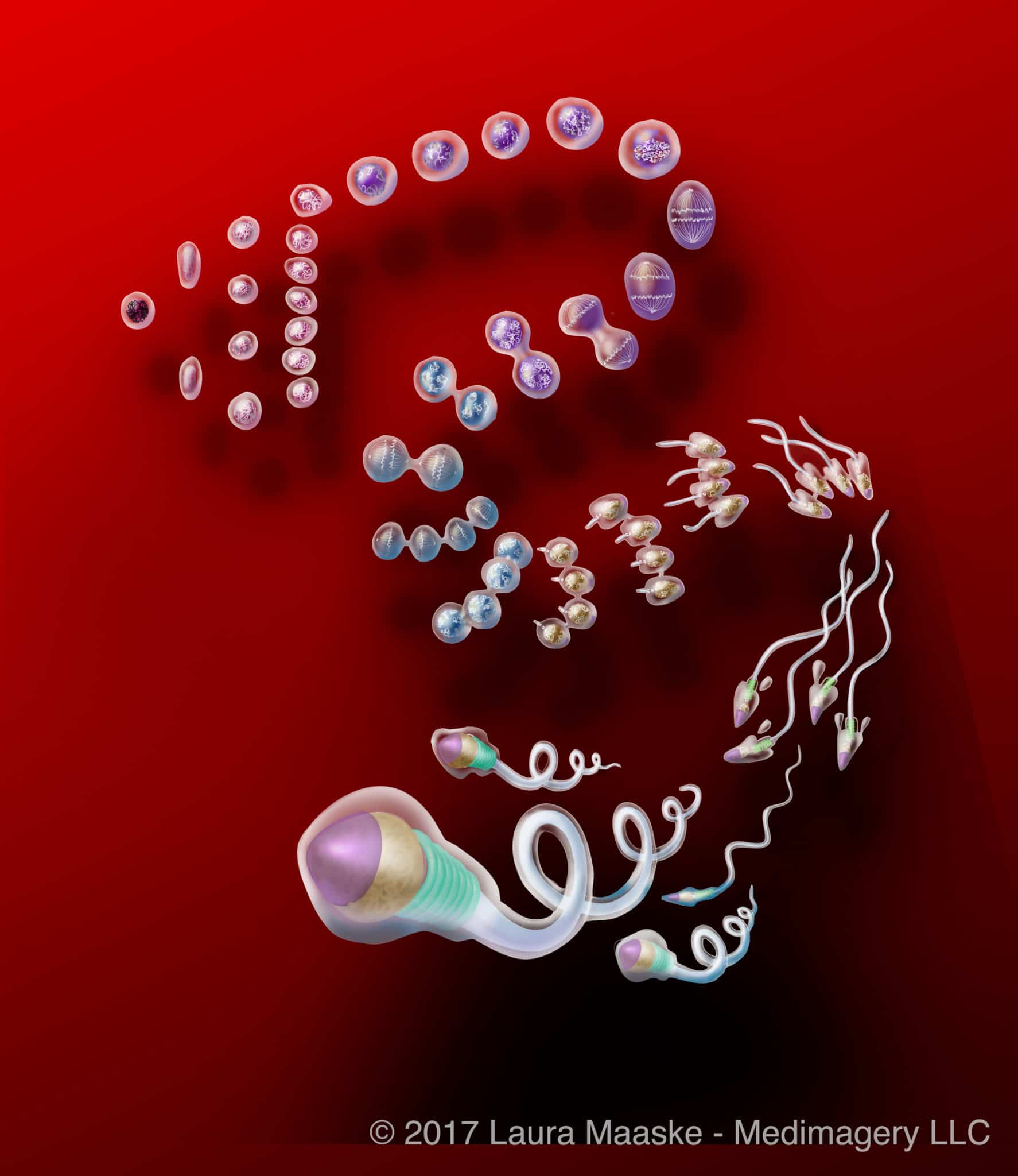
Primary Spermatocytes enter Meiosis 1: Prophase, Metaphase, Anaphase, and Telophase. In Prophase 1, 46 pairs of chromosomes attach to their complement pair of chromosomes, forming 23 tetrads. Chromosomes within each tetrad interchange genetic information. As the nuclear membrane begins to dissolve, centriole pairs begin to move to either pole of the cell. During Metopahase 1 the centriole spindles attach to chromosome pairs, which have lined up along the midline. In Anaphase 1 the centrioles pull chromosomes to either pole. By Telophase 1 each pole possesses the hapoid set, 23 pairs. A cleavage furrow develops, separating the chromosomes.
Haploid Secondary Spermatocytes
in Meiosis II
8 Hours Duration
Stage 3
Secondary Spermatocytes pass through Meiosis II, involving the same four steps as Meiosis 1: Prophase, Metaphase, Anaphase, and Telophase. Meiosis II resembles mitosis in that the 23 pairs of chromosomes do not exchange genetic information. They simply break from their pairs, becoming 23 single chromosomes which migrate to either pole. The product of Meiosis II is a group of four cells interconnected via intercellular bridges. Intercellular bridges synchronize division among the cells; without them the cells would perish. The product of Meiosis II is spermatids.
Diploid Spermatids
in Spermatogenesis
22 Days Duration
Stage 4
Spermatids first enter a Golgi Phase: granules fuse to form an ?acrosomal vesicle? on either side of the nucleus. On the other side, meanwhile, centrioles begin forming the sperm tail: one centriole attaches to the nucleus and the other forms at the anterior tail. During the Cap Phase, the acrosome vesicle begins spreading over the nucleus, now called the ?Acrosomal Cap?. During the Acrosome Phase the Acrosomal Cap, now ?Acrosome?, continues enveloping the nucleus. Mitochonndria form in a ?neck?. The nucleus condenses. Spermatids release nearly half their volume as residual cytoplasm, forming a more compact stremlined appearance.
Basic Development of Egg and Sperm
Egg and sperm are initially produced through a process called meiosis. Sperm are produced in the male testes. Cells with 46 chromosomes, called diploid cells, divide in such a way as to produce cells with 23 chromosomes, called haploid cells. The result is haploid sperm cells containing 23 chromosomes.

Spermatozoon, Fully Developed
In females, diploid cells, with 46 chromosomes, in the ovary, divide to produce haploid cells. These haploid cells are eggs, each of which contains 23 chromosomes.
Sperm migrate from the testes through and out the penis during sex. Eggs, one of which is produced each month, migrate from the ovary to the uterus once a month. Egg and sperm typically meet in the fallopian tube, and migrate to the uterus over a course of 5 days. Eggs which do not meet sperm are discharged at the time of a woman’s menstruation.
During a single ejaculation, a human male can release from 50 million to 1 billion spermatozoa. Typically, only one of these spermatozoa will achieve entry to an egg. When an egg cell becomes fertilized with a sperm cell, the two haploid cells unite and integrate their chromosomic material to become a diploid cell, a fertilized egg. The gender of the future baby is determined by the male genetic contribution, at the time of conception. The diploid cell is the first cell of a human body. It holds 46 chromosomes, half contributed by each of its parent cells. This 46 chromosome cell then divides by the mitotic process, mitosis, thus producing a two celled organism. Each of these calls divides and grows, thereby beginning the human life as a multicellular organism.

Meiosis in Human Female and Male Haploid Cells
Other Resources
Although I am an illustrator, I am still always looking for beautifully prepared animations and illustrations. This one is an evocative display of cell division. If you didn’t already have a sense of the dramatic life of a cell, you will after watching this:
Cell Division and the Cell Cycle
Onwards: Fetal Development
To further explore this topic, continue on with a fetal development series I illustrated here: Fetal Development Illustrated

November 20, 2013
Laura Maaske, MSc.BMC, Medical Illustrator & Medical Animator | e-Textbook Designer

[jp_post_view]

