Call Laura: +1 262 308 1300 | Email : laura@medimagery.com
Shadow Divider Down

Most Famous Medical Illustrator in History: Leonardo Da Vinci Anatomy

Shadow Divider Up

Most Famous Medical Illustrator in History: Leonardo Da Vinci Anatomy

Leonardo dissected more than 30 diseased and healthy human corpses during his lifetime, as a means of discovering

“Da Vinci had come to believe in Alberti’s advice that an artist should build a picture of the body from inside out. First conceiving of the skeleton then the skin then the clothes… He became typically obsessive as he carried forward Alberti’s injunction that artists should conceive a body from inside out. Leonardo wrote, ‘It is necessary for the painter in order to be good at arranging the parts of the body in attitudes and gestures which can be represented in the nude to know the anatomy of the sinews and bones and muscles and tendons.’”

Walter Isaacson, Leonardo Da Vinci

Most Famous Medical Illustrator in History: Leonardo Da Vinci Anatomy

Leonardo Da Vinci’s Anatomical Drawings

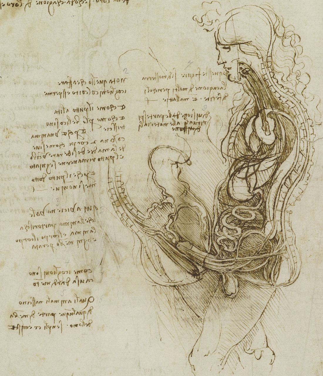
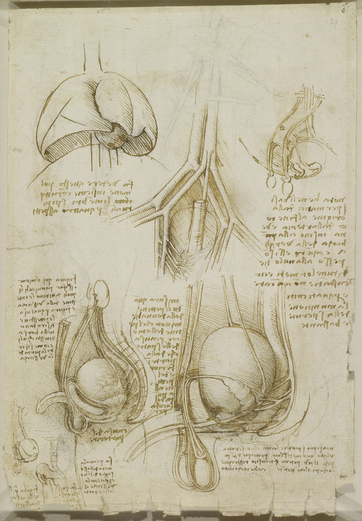
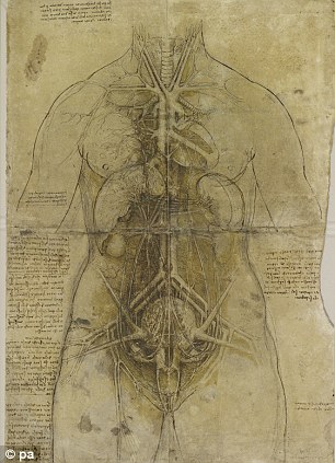
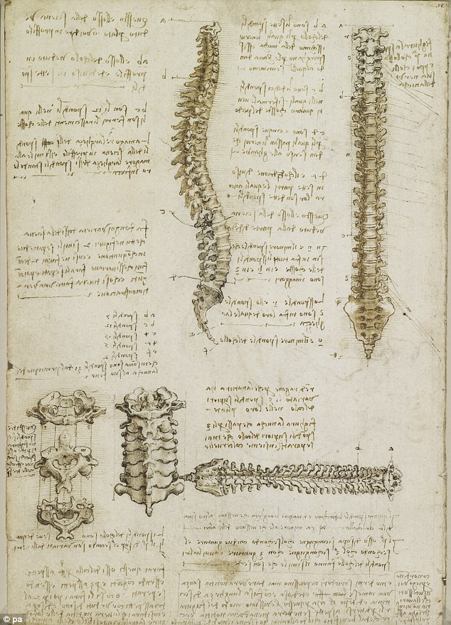
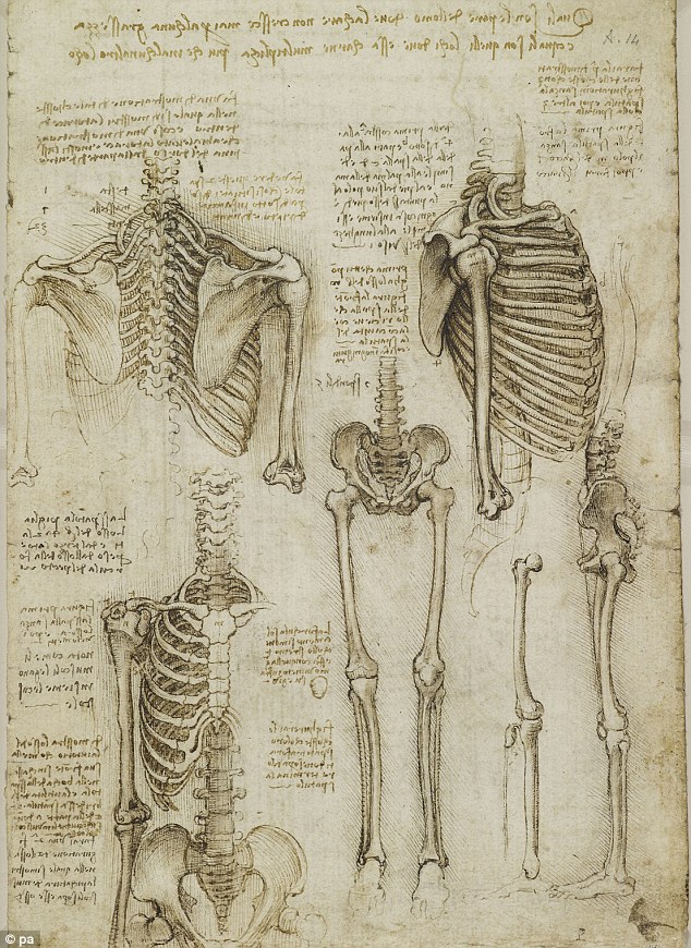
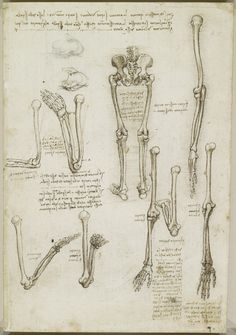
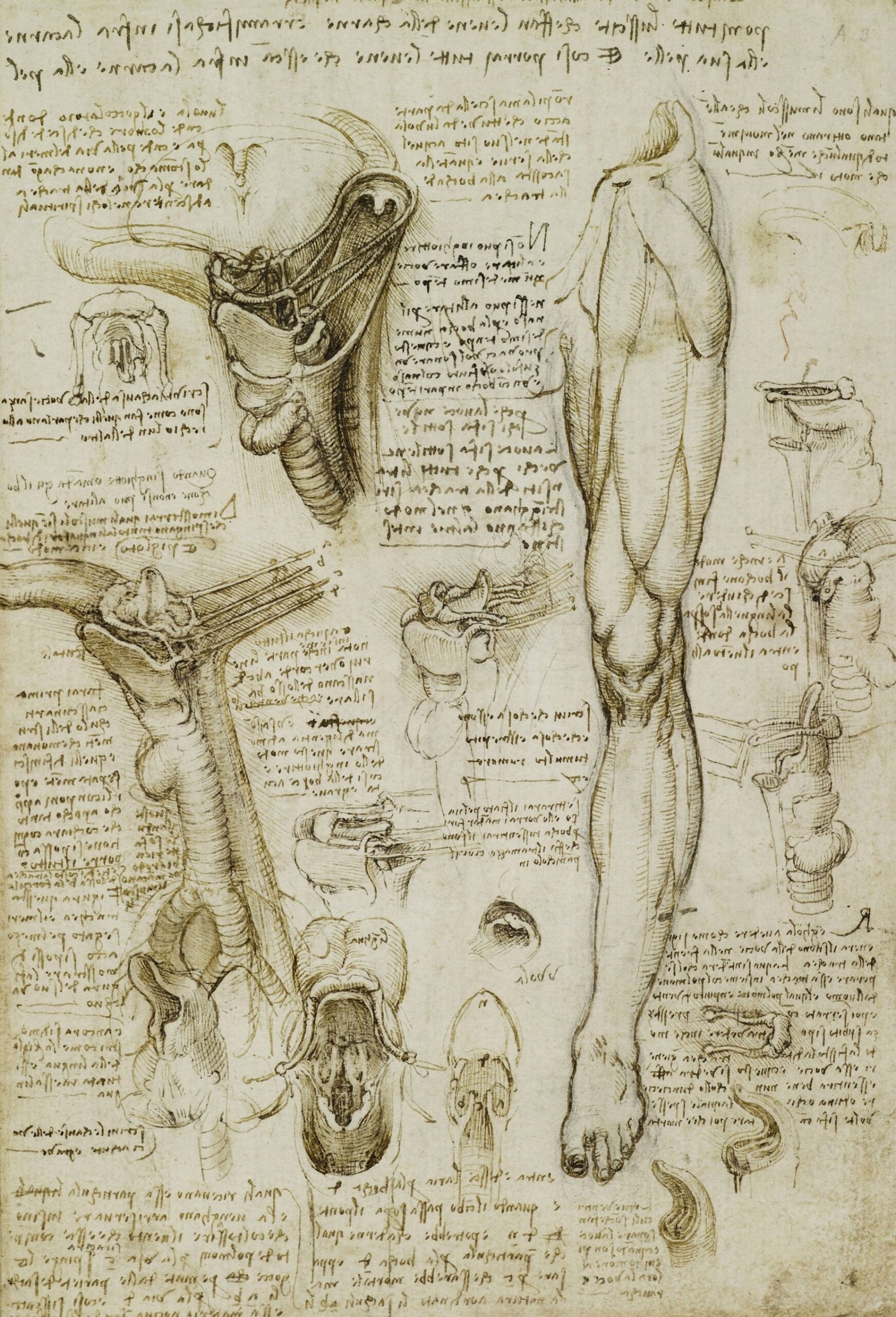
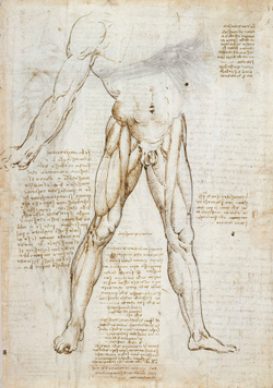
Leonardo’s interest in anatomy began when he was working for the Milanese benefactor, Ludovico. April 2, 1489. His first drawing was a skull. However, because it was illegal and considered sacrilegious to dissect the human body, Da Vinci, while curious about anatomy, did not often have the chance to study anatomy first hand. In the course of his life, he dissected about 30 diseased and healthy human corpses, as a means of discovering human anatomy. Philosophers such as Plato and Aristotle were curious about the body. But they did not dissect it in order to create a realistic model for reference. Consequently, Da Vinci’s renderings are remarkably accurate

Leonardo Da Vinci recorded his careful observations of the human body in a notebook called, “Book entitled On the Human Figure”. On one page he drew his most notable drawing, The Vitruvian Man. The Vitruvian Man depicts a man standing in the middle of a circle and a square, showing two different poses. Da Vinci intended for these notebooks to be used in the medical field.

None of Leonardo’s astute observations or drawings were ever published during his lifetime due to the taboo about dissecting the body. His notebooks were not really known until 250 years after his death or popular until 1965.

The following is a collection of Leonardo DaVinci anatomical drawings, including illustrations related to anatomy, relevant to what a medical illustrator might study in order to become a better medical artist. I have even included a few examples of animal anatomy.

Leonardo da Vinci 1 January 1489


https://i0.wp.com/medimagery.com/wp-content/uploads/2018/04/View_of_a_Skull_III.jpg?resize=434%2C455

”View of a Skull”’ (c. 1489) is a drawing by Leonardo da Vinci.
Source: http://www.visi.com/~reuteler/leonardo.html {{pd}}Category:Leonardo da Vinci


https://i0.wp.com/medimagery.com/wp-content/uploads/2018/04/View_of_a_Skull_II.jpg?resize=464%2C438

”’View of a Skull”’ (c. 1489) is a drawing by Leonardo da Vinci.
Source: http://www.visi.com/~reuteler/leonardo.html {{pd}}Category:Leonardo da Vinci


https://i0.wp.com/medimagery.com/wp-content/uploads/2018/04/Leonardo-skull.jpg?resize=568%2C534

Study of a skull by Leonardo da Vinci between 1510 and 1511

https://i0.wp.com/medimagery.com/wp-content/uploads/2018/04/Leonardo_da_vinci2C_Studies_of_human_skull.jpg?resize=651%2C962

Studies of human skull 1489


https://i0.wp.com/medimagery.com/wp-content/uploads/2018/04/1240px-Skull_in_profile_to_right_MET_DP823728.jpg?resize=1239%2C1024

Da Vinci’s Skull in profile to right 1645. Etching; only state


https://i0.wp.com/medimagery.com/wp-content/uploads/2018/04/1115px-Cross-section_of_skull_in_profile_view2C_showing_the_intercra_Wellcome_V0007777EL.jpg?resize=1115%2C1024

Cross-section of skull in profile view, showing the intercranial nerves and vessels. Da Vinci’s Etching by Wenceslaus Hollar after Leonardo da Vinci, 1651. Iconographic Collections


Da Vinci nerves of the brain, cranial nerves and other brain structures

Da Vinci nerves of the brain, cranial nerves and other brain structures


Related image

Related image


Human Cardiac Anatomy by Leonardo Da Vinci

Human Cardiac Anatomy by Leonardo Da Vinci


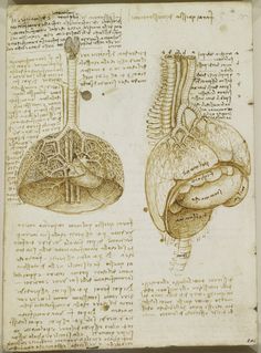
The 500 year old drawing above is Leonardo da Vinci’s description of the heart he dissected of a recently deceased 100 year old man. It is the first known description of cardiac disease, in which he noted that the arteries may be clogged if they collect debris. Da Vinci also drew the valves of the heart and noted how blood flowed through the aorta to the heart and how with this pulse, the valves opened and closed by way of currents in the blood and the strength, and how blood flowed through the heart as a result of these currents. His observations were so accurate that only with modern MRI technology did the genius of his observations become clear. Da Vinci also observed that the heart is a muscle and that it does not warm the blood, which was not well understood at the time. He noted the four chambers of the heart and that the heart beat of the left ventricle is related to the pulse of the wrist. Da Vinci explored not only the human heart, but also the pig heart.


card

Cardiac Muscle by Leonardo Da Vinci


Drawing of external genitalia and vagina, with notes; notes on the anal sphincter and diagrams of suggested arrangement of its fibers and its mode of action. Royal Collection © Her Majesty Queen Elizabeth II

Drawing of external genitalia and vagina, with notes; notes on the anal sphincter and diagrams of suggested arrangement of its fibers and its mode of action. Royal Collection © Her Majesty Queen Elizabeth II


Da Vinci Human Fetus

Leonardo da Vinci, The Foetus in the Womb. Pen and ink over red chalk, circa 1510-1513. Royal Collection, The Windsor Castle


Leonardo da Vinci's sketches of a fetus in the womb, made between 1510 and 1513.

Leonardo da Vinci’s sketches of a fetus in the womb, made between 1510 and 1513.

Related image

Dedicated: Da Vinci dissected more than 30 human corpses to learn about the human anatomy so his paintings and sculptures would be as realistic and accurate as possible


Dedicated: Da Vinci dissected more than 30 human corpses to learn about the human anatomy so his paintings and sculptures would be as realistic and accurate as possible


Image result for Leonardo da Vinci, The Bones and Muscles of theShoulder. Pen and ink with wash over black chalk, circa1510-1511.Royal Collection, The Windsor Castle


Master of the arts: Many professors of anatomy say they still use da Vinci's drawings to teach medical students as the accuracy of his work has rarely been surpassed


Detailed: Da Vinci's sketches of muscles and skeletons foreshadow modern techniques, such as MRI scans and 3D computer modelling, to 'an astonishing degree'

Related image

Detailed: Da Vinci's sketches of muscles and skeletons foreshadow modern techniques, such as MRI scans and 3D computer modelling, to 'an astonishing degree'


Muscles of the arm Da Vinci

Muscles of the arm, Da Vinci


Related image

Eerie: The notebooks, which date between 1452 and 1519, contain incredibly detailed images of the human body


Da Vinci shoulder anatomy

Da Vinci shoulder anatomy


Three studies, one on a larger scale, of a man’s right arm and shoulder, showing muscles; three studies of a right arm; a diagram to illustrate pronation and supination of the hand. Royal Collection © Her Majesty Queen Elizabeth II

Three studies, one on a larger scale, of a man’s right arm and shoulder, showing muscles; three studies of a right arm; a diagram to illustrate pronation and supination of the hand. Royal Collection © Her Majesty Queen Elizabeth II


Da Vinci arm anatomy


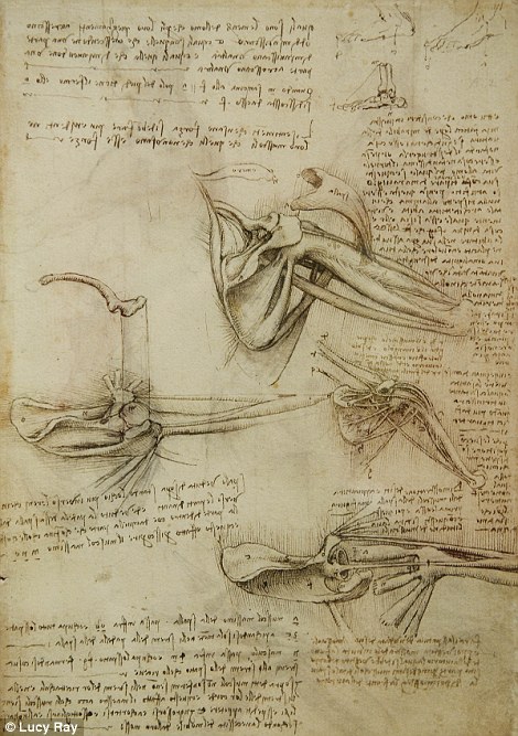
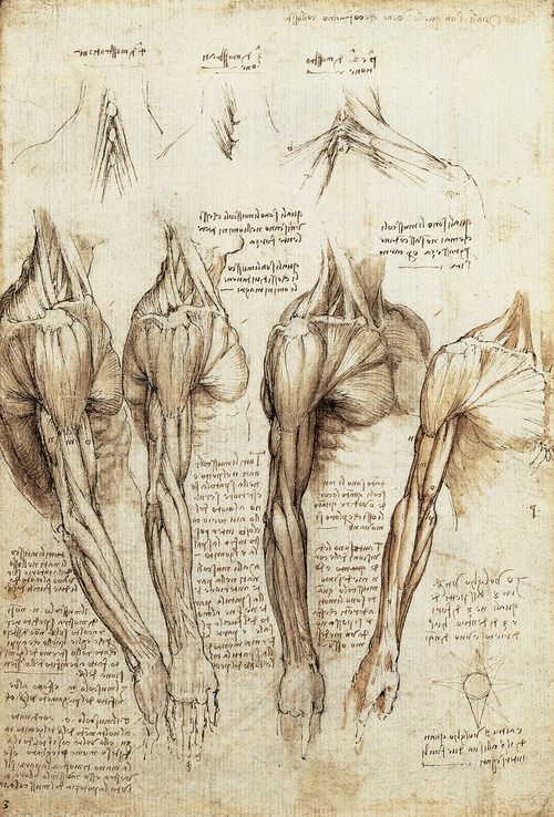
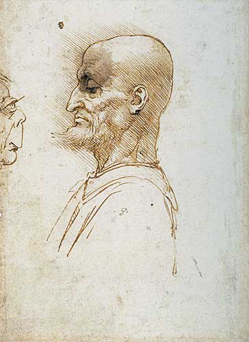
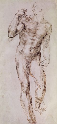
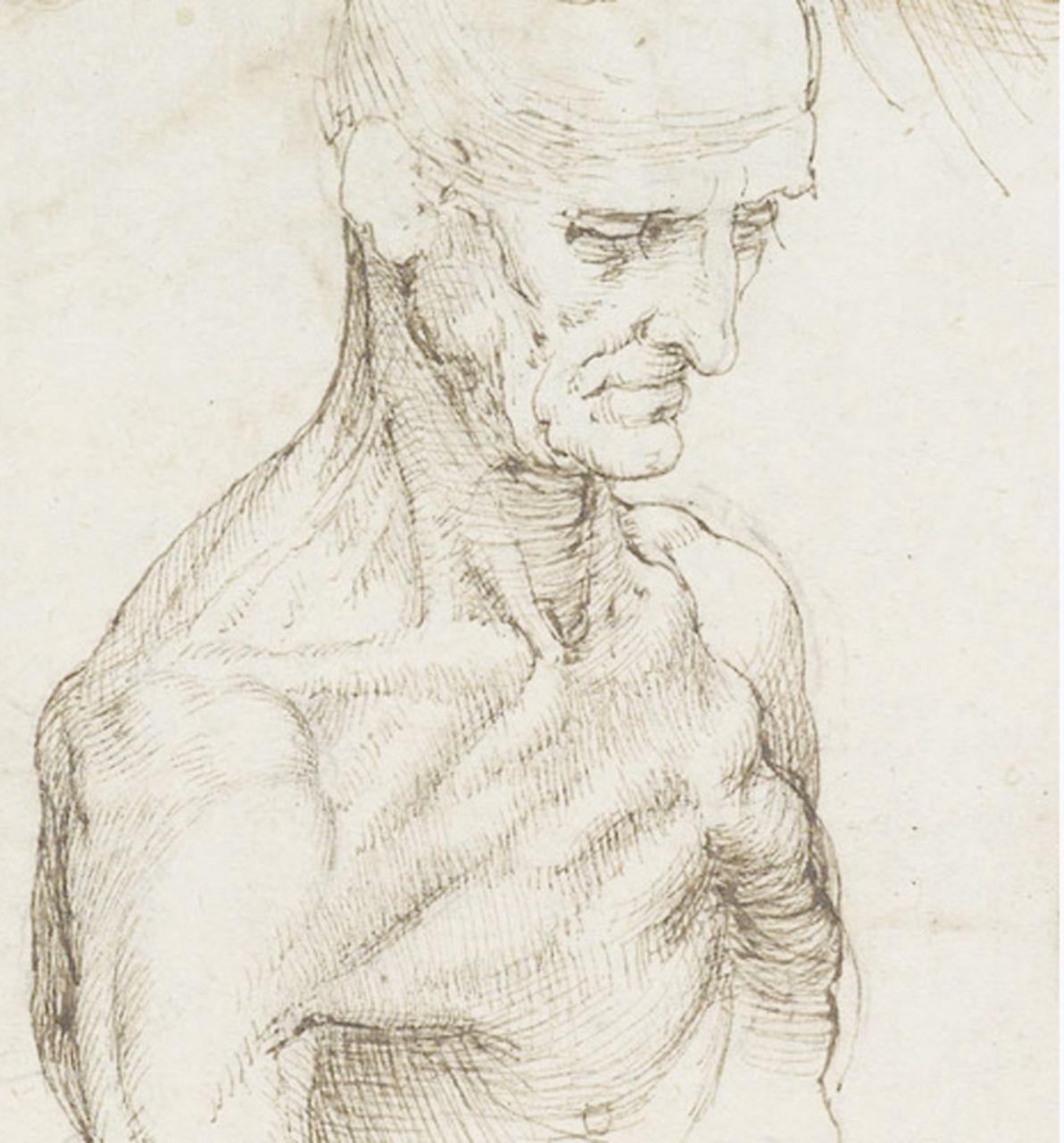
Leonardo da Vinci, The Bones and Muscles of the Shoulder. Pen and ink with wash over black chalk, circa 1510-1511. Royal Collection, The Windsor Castle

human arm bones by Leonardo Da Vinci

Human arm bones by Leonardo Da Vinci


da Vinci Hand Anatomy

da Vinci Hand Anatomy


Image result for Leonardo da Vinci, The Bones and Muscles of theShoulder. Pen and ink with wash over black chalk, circa1510-1511.Royal Collection, The Windsor Castle
Related image

Related image

 A study of a man’s left leg, stretched forward; beside it, a man’s legs seen from behind; below is a man standing, turned in profile to the left, with his left leg advanced; to the right are two studies of the bones of human left legs and thighs, and one of an animal; with many notes on the muscles. Royal Collection © Her Majesty Queen Elizabeth II

A study of a man’s left leg, stretched forward; beside it, a man’s legs seen from behind; below is a man standing, turned in profile to the left, with his left leg advanced; to the right are two studies of the bones of human left legs and thighs, and one of an animal; with many note


Five studies of the bones of the leg and foot; a drawing of the knee joint and patella; two studies of the bones of a right leg with the knee flexed; the muscles of a right buttock, thigh and calf. Royal Collection © Her Majesty Queen Elizabeth II

Five studies of the bones of the leg and foot; a drawing of the knee joint and patella; two studies of the bones of a right leg with the knee flexed; the muscles of a right buttock, thigh and calf. Royal Collection © Her Majesty Queen Elizabeth II


Da Vinci’s Love of the Rational Human Form


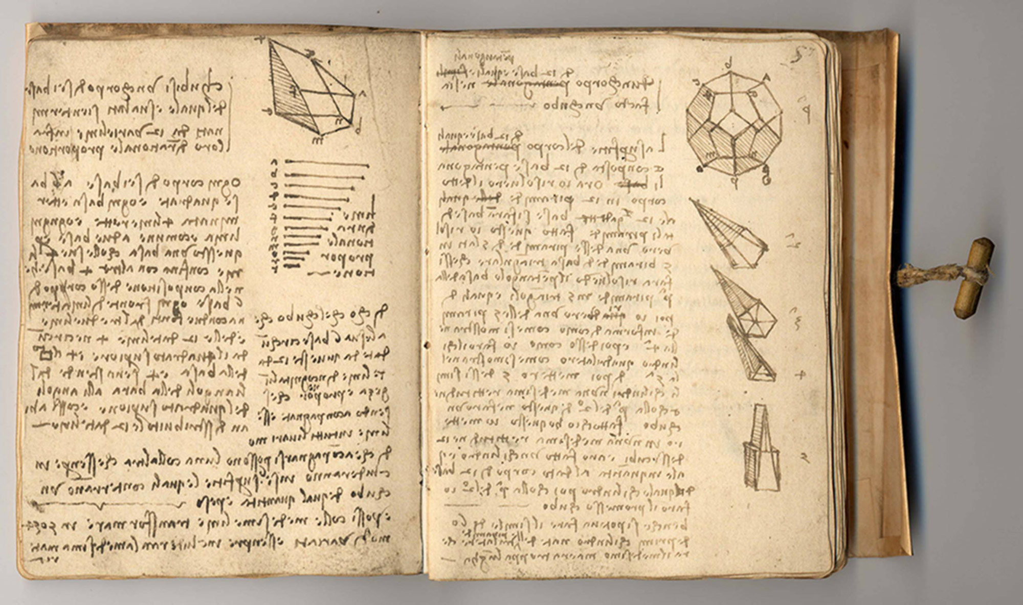
Leonardo Da Vinci made extensive, almost obsessive measurements of the human form, noting thousands of ratios between various body parts in various standing and sitting positions. To see a beautifully documented description of his observed proportions, visit the Artist’s Network page on Da Vinci proportions, an analysis very thoroughly explored, and a direct translation of Da Vinci’s notes.


Vitruvian Man


Vitruvian Man. drawn in 1490, is s special example of Da Vinci and his time. Da Vinci, a true Renaissance man, explored many fields as a sculptor, painter, writer, inventor, mathematician, engineer, architect, and anatomist. It is the view at this time that all natural phenomena can be explained with math. And while the principle is remarkably true. It also proved untrue in some remarkable ways, such as Galileo forcing a geometry on the movement of planets. , though not always accurate, to represent the human body, an inherently non-formulaic or mathematically precise form, and render it in a mathematical way. Here is a great video to explain this perspective.


While Vitruvian man is not a precise representation of how the human form behaves, we still find it beautiful. And Da Vinci’s effort to render the world in a predictably proportioned way is one of the core aspirations of his anatomy notebooks. These notebooks are filled with notes on proportion.

https://i0.wp.com/medimagery.com/wp-content/uploads/2018/04/Keplers_solar_system.png?resize=755%2C923

Keplar’s model of a universe, explained with geometric forms. The model did not fit the actual behavior of the planets. But it was one example of the Renaissance passion for math


“The man who blames the supreme certainty of mathematics feeds on confusion, and can never silence the contradictions of sophistical sciences which lead to an eternal quackery.” -Da Vinci

“There is no certainty in sciences where one of the mathematical sciences cannot be applied, or which are not in relation with these mathematics.” – Da Vinci


Study of the proportions of the head.

Francesco Melzi – Portrait of Leonardo – WGA14795


Two anatomical studies


Related image
Image result for Leonardo da Vinci, The Bones and Muscles of theShoulder. Pen and ink with wash over black chalk, circa1510-1511.Royal Collection, The Windsor Castle


Natural and Scientific Illustrations and Explorations of Da Vinci

Although I did not find an example of Da Vinci


Image result for Leonardo da Vinci, The Bones and Muscles of theShoulder. Pen and ink with wash over black chalk, circa1510-1511.Royal Collection, The Windsor Castle
A Rider on a Rearing Horse. Leonardo da Vinci (Italian, 1452-1519). Metalpoint reinforced with pen and brown ink on a pinkish prepared surface. 141 mm x 119 mm. Not before 1481.

A Rider on a Rearing Horse. Leonardo da Vinci (Italian, 1452-1519). Metalpoint reinforced with pen and brown ink on a pinkish prepared surface. 141 mm x 119 mm. Not before 1481.


Leonardo da Vinci (Italian, Vinci A Bear Walking

Leonardo da Vinci (Italian, Vinci A Bear Walking


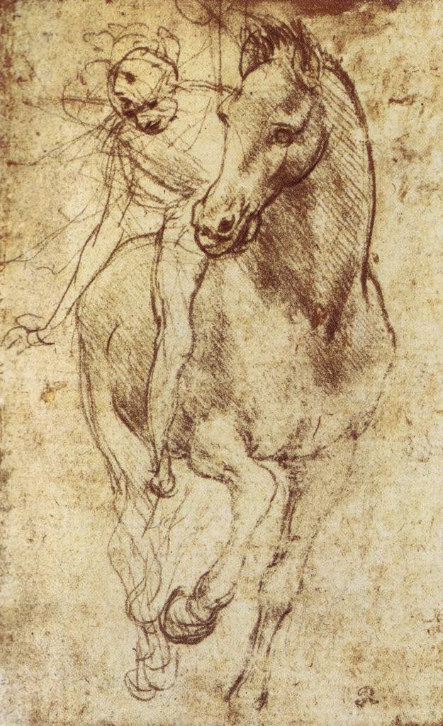
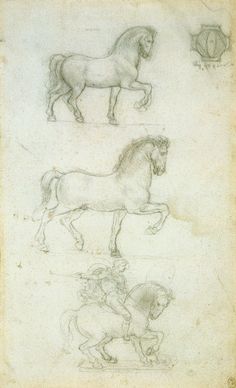
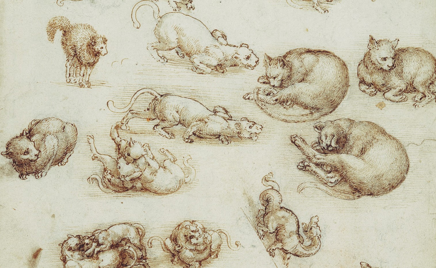
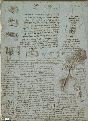
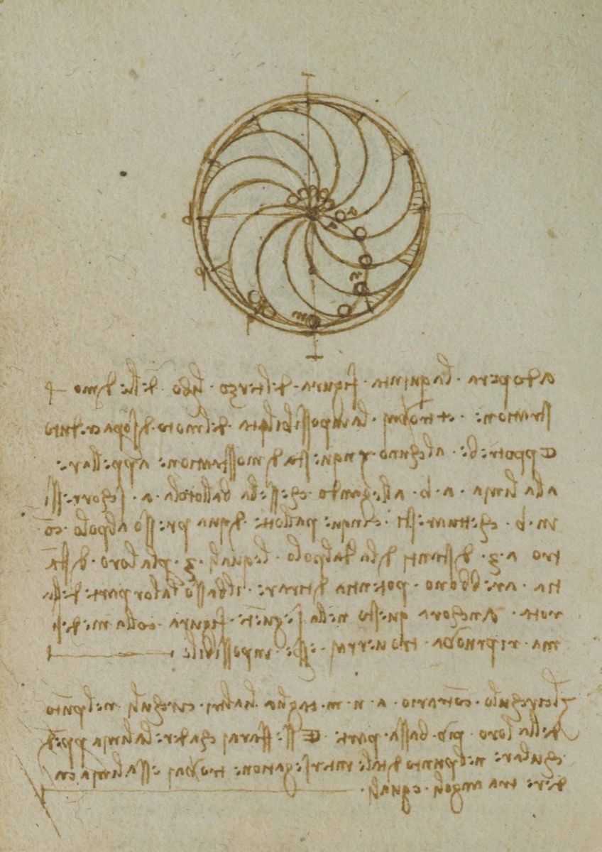
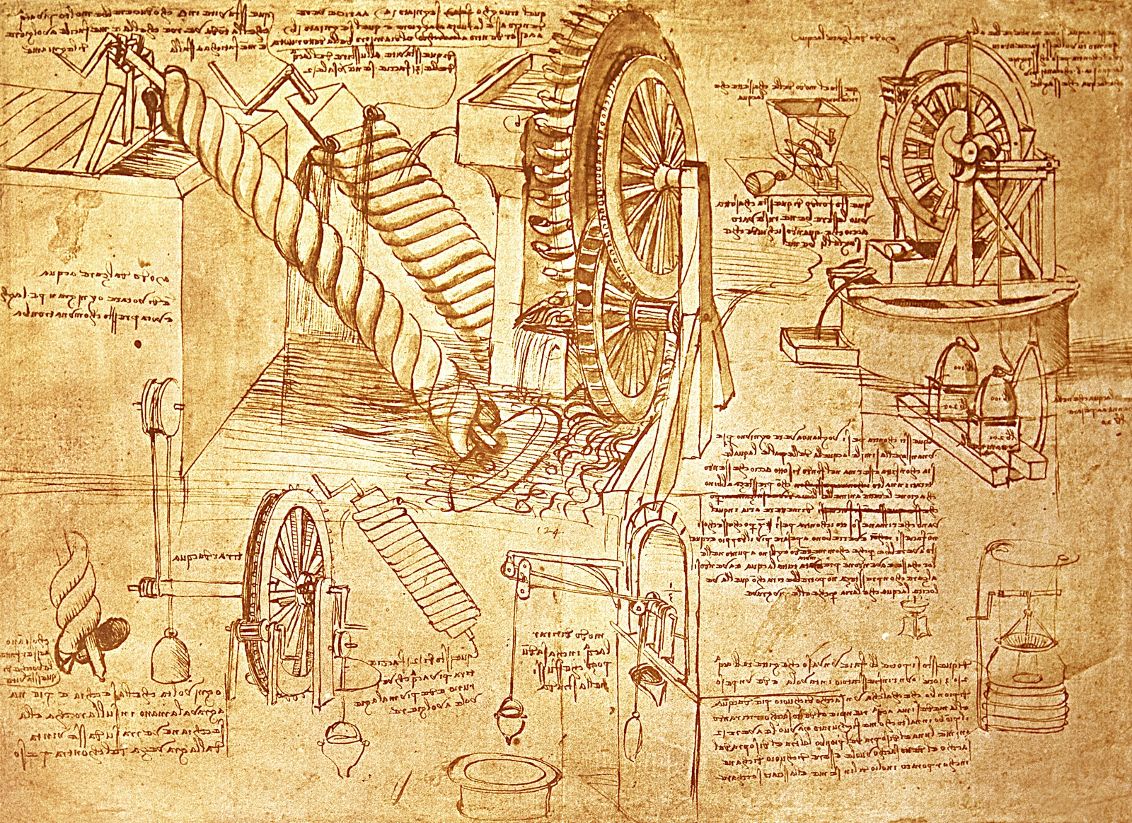
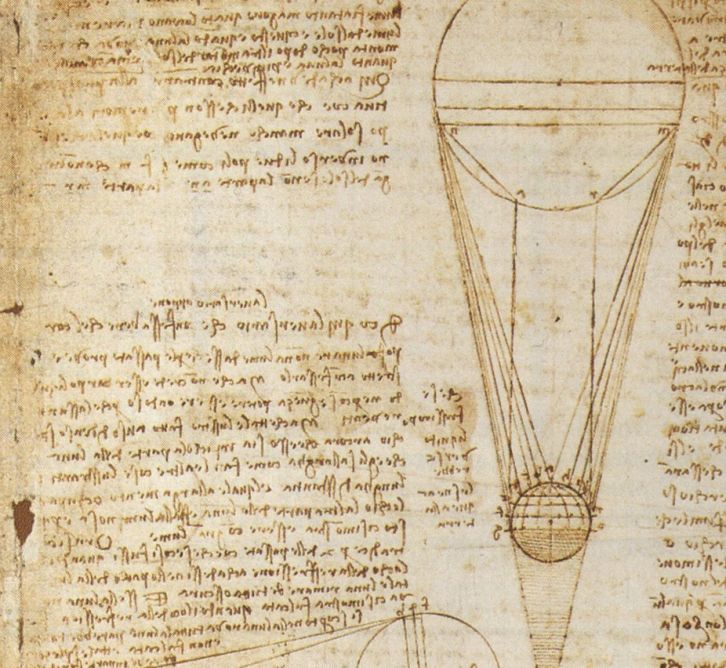
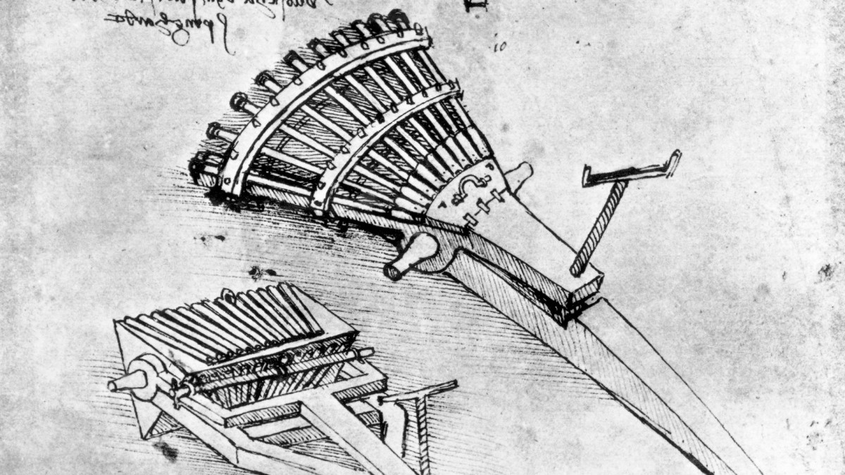
Da Vinci’s Anatomy of Objects as related to His Anatomical Explorations

These Illustrations show Da Vinci’s love of mechanics. I included them in my human anatomy collection because they reveal a love of the “inner workings” of objects. This is the same love that a medical illustrator embraces in everyday drawing. And I believe Da Vinci’s love of anatomy go hand in hand with this curiosity for mechanics:


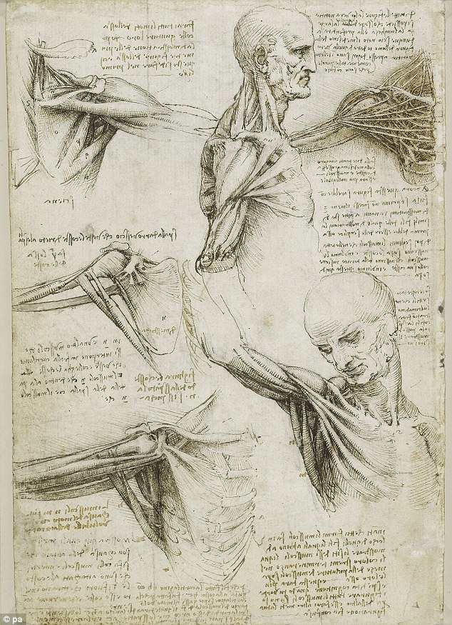
As a great artist, Leonardo had two advantages over his contemporary anatomists. First of all, as a sculptor, engineer, architect, he had an intuitive understanding of form — when he dissected a body, he could understand in a very fluid way how the different parts of the body fit together, worked together. And then, having made that understanding, as a supreme draftsman, he was able to record his observations and discoveries in drawings of such lucidity, he’s able to get across the form, the structure to the viewer in a way which had never been done before and, in many cases, has never been surpassed since.

Leonardo intended to publish his drawings as an illustrated treatise on human anatomy, but when he died in 1519, his anatomical papers were buried amongst his private possessions and vanished from public sight. In the early 1600s, around 600 of his surviving drawings were bound in a single collection and by the end of the century, they mysteriously made their way to the Royal Collection. Leonardo da Vinci: Anatomist gathers 90 of these seminal drawings, contextualized in a discussion of their anatomical significance. Accompanying the books is an iPad app, presenting 268 pages of Leonardo’s notebooks in magnificent high resolution.

Nature Video was invited to Windsor Castle to see some of Leonardo da Vinci’s anatomical drawings. The drawings show that Leonardo did more than dabble in the sciences; he carried out experiments and made staggering medical discoveries which could have transformed the study of anatomy in Europe — had they not languished unpublished for centuries. In this video, Senior Curator Martin Clayton shows us three of Leonardo’s most intriguing anatomical studies. The drawings in this video will be part of an exhibition at The Queen’s Gallery in Buckingham Palace from 4 May to 7 October 2012 http://www.royalcollection.org.uk/exh… Martin has also written a commentary for Nature’s Books & Arts section: Leonardo’s anatomy years http://www.nature.com/uidfinder/10.10…


_____________________________________


April 20, 2017

Laura Maaske, MSc.BMC.

Biomedical Communicator

Medical Illustrator

Medical Legal Illustrator

Medical Animator

Health App Developer

medimagery_medical-illustrator-350
_____________________________________
Laura Maaske – Medimagery LLC
Medical Illustration & Design


Pin It

About Laura

Medical Illustrator; science and art lover.