Medical Illustrations of Abdominal Organs & Digestive System
The Digestive System Has Two Main Functions:
- Convert food into nutrients your body needs
- Rid the body of waste
Main Contents of the Digestive System
The human body has specifically evolved to suit the available nutrients in the environments in which humans evolved. And there are several specialized organs nature has designed to make the most of ingested food.
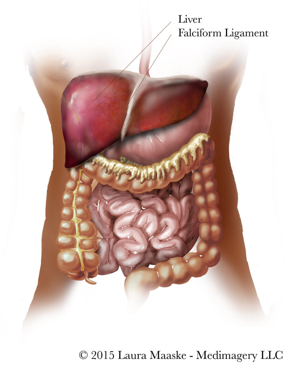
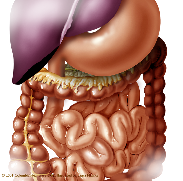
The human abdominal cavity stretches from the diaphragm to the pelvis at the pelvic brim and contains the hollow and tube-like alimentary tract, or digestive organs, of the body. These vital organs include the mouth, esophagus and stomach. The stomach continues into the small intestine, which consists of the duodenum, jejunum, ileum, and cecum. Digestion continues with the large intestine: ascending (including the appendix), transverse colon, descending colon, sigmoid colon, and the rectum. There are many accessory organs, solid in structure, which aid in digestion and these include the liver, gall bladder, kidneys, pancreas, and spleen.

Mouth & Stomach
As food is swallowed it passes through the esophagus by a wavelike movement called peristalsis, to the stomach. Enzymes, including proteases, amylases, and lipases released throughout the digestive system, break down the swallowed food. Even the mouth releases amylase and lipase in the saliva. Amylases break down carbohydrates, lipases break down fats, and proteases break down proteins. The stomach and small intestine release mainly proteases in their exudate.

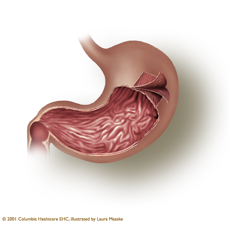
This gastric illustration depicts the three layers of smooth muscle which line the stomach wall.
Smooth muscle in the stomach contracts during digestion, in order to break down nutrients. The muscle layers in the human stomach are each striated in different directions. Because smooth muscle can only contract in one direction, it is functionally adaptive for the stomach, which must contract as an organ overall, to contract in three directions. Another muscle in the body which has muscle fibers running in different directions is the tongue. The stomach is the only organ in the digestive system to have three muscle layers. The rest of the gastrointestinal tract, or GI tract, contains only two muscle layers.

Small Intestine
The small intestine is roughly 22 ft long (7 meters), and one inch wide (2.5 cm). And because of the microvilli which make a very convoluted surface, the actual surface area of the small intestine is 2,700 square ft (250 square m). This is roughly the same square surface of a tennis court!
Large Intestine
The large intestine gets its name because it is wider than the small intestine. But in fact, it is also the “short intestine”, as it is far shorter than the small intestine. The large intestine is only 25% as long as the small intestine. It’s major function is
- to conserve water by extracting liquid from food and returning it to the body.
- Convert the remaining food to feces before releasing it by defecation.
The large intestine includes the cecum, colon, rectum, and, by some viewpoints, the anal canal.
Rectum
The rectum is the last section of the digestive system. It holds and forms the feces before it is released.
Accessory Organs to the Digestive Tract
- Gall bladder – assists in lipid digestion and concentrates bile produced by the liver.
- Kidneys – regulate enzymes, maintain acid and salt balance, and regulate of blood pressure and water balance. The kidneys remove waste and divert toxins to the ureter and urinary bladder. The kidneys produce urine, which contains urea and ammonium. The kidneys also manufacture the hormones erythropoietin and calcitriol; and the enzyme renin.
- Pancreas – a digestive organ as well as an endocrine gland. As a digestive organ it releases pancreatic enzymes for the small intestine to break down proteins, lipids, and carbohydrates. As a gland it produces vital hormones like insulin, glucagon, pancreatic polypeptide, and somatostatin.
- Spleen – as part of the lymphatic and immune system, and less vital than the other organs, the spleen filters blood. It removes the old red blood cells from blood and holds a reserve of blood if there should be sudden hemorrhage somewhere in the body. The spleen recycles iron, removes bacteria which have been identified as foreign by immune cells, and synthesizes antibodies.
Other Contents of the Abdomen
Generally, the bladder and ureters, uterus and ovaries, are considered part of the pelvis and not the abdomen, due to the perimeter of the abdominal wall itself, the peritoneum. However, there is some disagreement about this. These organs are considered to be in a retroperitoneal position. A retroperitoneal position is to be located in the back of the peritoneal or pelvic cavity as opposed to within the abdominal cavity.
Blood Supply to the Digestive Tract
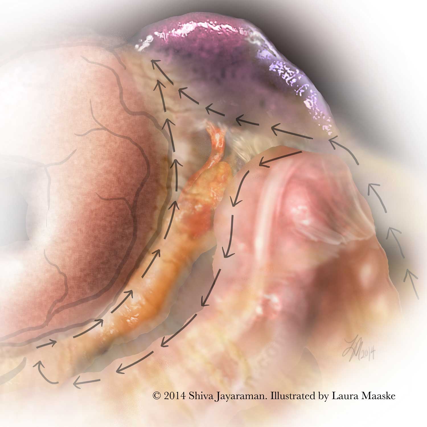
The colon receives fresh arterial blood on the right side of the body from the body from the superior mesenteric artery (SMA), and on the left side from the inferior mesenteric artery (IMA). The left gastric artery supplies the lesser curve of the stomach. And the lft and right gastroepiploic arteries supply the greater curve of the stomach. The splenic artery supplies the spleen with blood. The left and right hepatic arteries supply the liver. The right and left renal arteries supply the kidneys.
Venous architecture is similar in structure to the colonic arterial supply. The liver receives drainage from the hepatic portal vein. The hepatic portal vein receives blood from the inferior mesenteric vein, superior mesenteric vein, and splenic vein.
The Mesentery: New Findings in the Anatomy of the Digestive System
The mesentery is a double layer of soft, fatty peritoneum that attaches the stomach, small intestine, pancreas, spleen, and other organs to the posterior wall of the abdomen, and lays over them like a blanket as a lining to the peritoneum.
An Irish surgeon, Prof J Calvin Coffey, of the University of Limerick, recently clarified the anatomical relationship of the mysterious mesentery. He revealed it as having a unified structure, more than an accessory of the large intestine. Previously it was thought to be a series of separate components. His research is published in the Lancet Gastroenterology & Hepatology. Mesenteric scientists are still unable to clearly define the function of the mesentery, but it may play a role in inflammatory bowel disease, colorectal cancer, diabetes, and obesity.

Perspective and Future Medical Illustrations: the Digestive System
The digestive system is an extraordinarily complex system which, despite our long historical search for understanding, still reveals to humans its subtle functions. This article is a very brief overview. How its anatomy relates to the structure of nutrient use and the immune system is an area currently under study. While I have not yet been asked to create illustrations to reflect the new research on the bacterial makeup of the digestive system and its impact on wellness, I hope to offer new information and research here in the future.

Medical Illustration of Abdominal Organs
Thank you for reading and feel free to offer comments or clarification relevant to your area of specialization.
Laura
May 11, 2017
Laura Maaske, MSc.BMC. Biomedical Communicator

Text Copyright © 2017 Medimagery – Laura Maaske LLC
Illustration © 2000 Columbia Healthcare EHC, illustrated by Laura Maaske – Medimagery LLC.
[jp_post_view]


1 Comment