
Liver Resection Illustrations: Laparoscopic Left Lateral Liver Sectionectomy
Liver Resection Surgical Illustration Series
Written and illustrated by Laura Maaske, MSc.BMC, Medical Illustrator & Medical Animator
What follows is a description and illustration series I created for the “Liver Resection Illustrations: Laparoscopic Left Lateral Liver Sectionectomy” surgical procedure, as performed by Dr. Shiva Jayaraman at the St. Joseph’s Health Center, Toronto, Canada,
When a surgical client asks me to illustrate a procedure, or to create surgical illustrations, it is because the surgeon has a new or innovative approach to the procedure. My aim is to create illustrations that reflect their personal contributions or adaptations of a specific surgical technique. This illustration series depicts a surgical procedure which is performed by Dr. Jayaraman at St. Joseph’s Health Care Center, Toronto. The procedure is performed to remove a tumor, and most typically in adults.
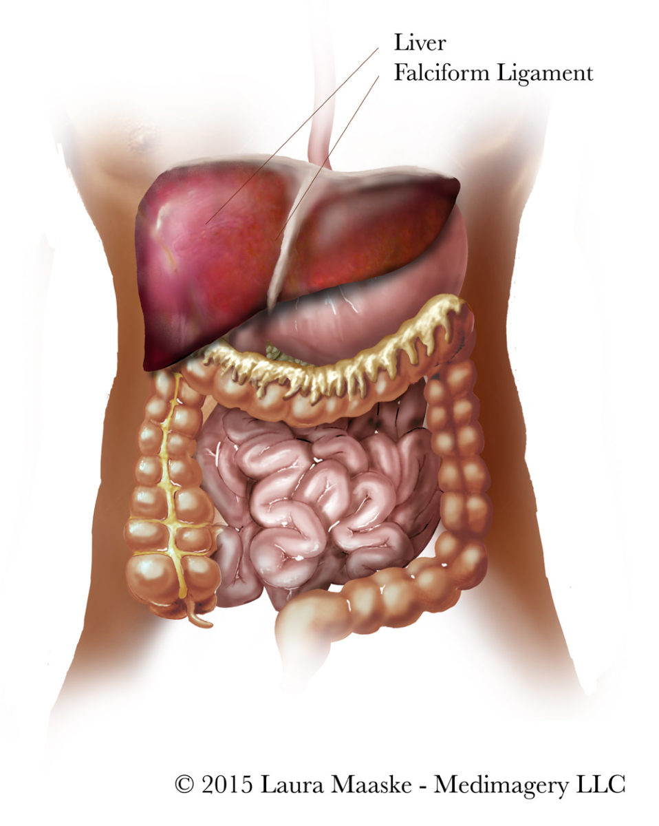
If you are reading this liver sectionectomy illustrations article because you did a search on the topic, you may already know the abdominal anatomy. Note below the falciform ligament. The falciform ligament is a remnant of the umbilical vein, which, during a human’s early development, delivers nutrients to the fetus. There are two components of the falciform: (1) a superficial remnant as you see on the drawing below, and a (2) deep component which is the round ligament (or ligemantum teres).
What is a Laparoscopic Left Lateral Liver Sectionectomy?
Laparoscopic Left Lateral Liver Sectionectomy is a surgical resection procedure. The left lateral section of the liver, or left lobe is removed: both segments 2 and 3. Note this anatomy in the illustration below. This includes all the liver tissue left of the falciform ligament.
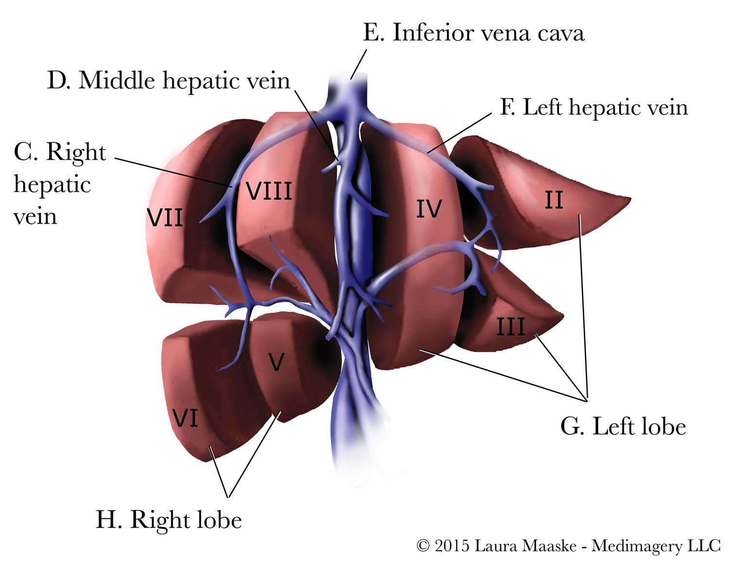
Surgeons use the falciform ligament during the procedure as an anatomical landmark, following it in dissection as its two layers separate and pass through and over the liver. This ligament condenses into to the left triangular ligament behind the liver, which in turn connects to the the underside of the diaphragm.
Steps of the Laparoscopic Left Lateral Liver Sectionectomy Procedure
Steps involved in the resection procedure are illustrated as follows:
1. Patient Positioning
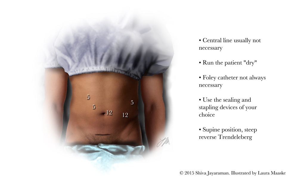
Note the surgical ports through which the laparoscopic instruments enter the abdomen.
2. Releasing the Falciform Ligament
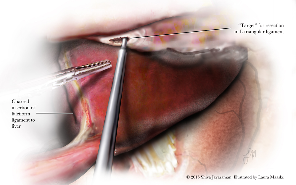
The falciform ligament is cut and released along the surface of the liver. Note the partially attached left triangular ligament with a little hole for target.
3. Opening Liver with Ultrasonic Shears
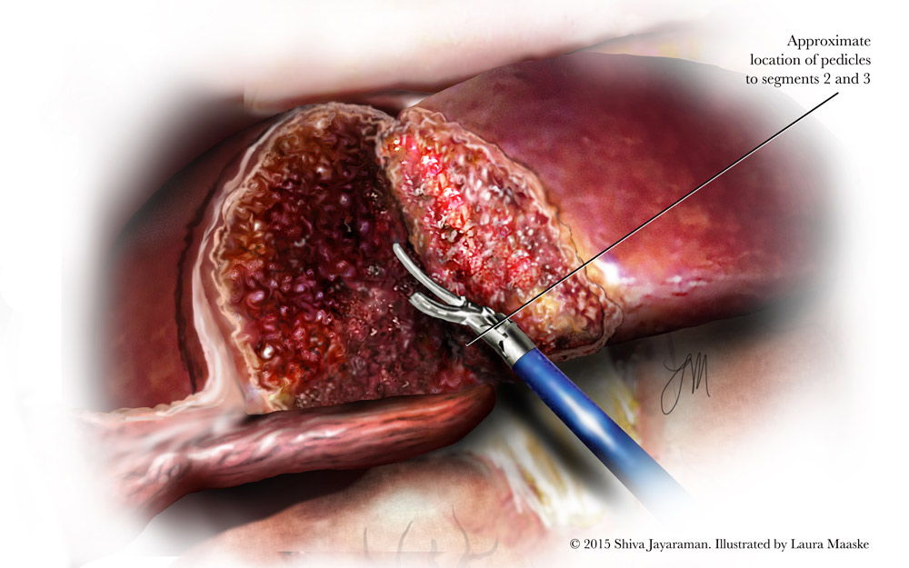
Ultrasonic shears are used to open up and begin separating the segments of the liver which will be resected carefully to bring the dissection up to the level of the pedicle.
4. Three Surgical Staples
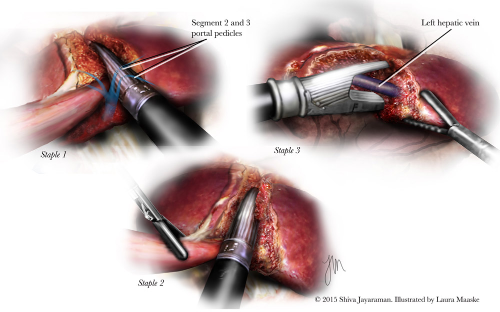
Three staples are used to stabilize the liver and prevent bleeding. Staple #1 goes over the pedicle for segments 2 and 3. Staple #2 is placed over some little branches of the hepatic veins, mostly going over some parenchyma between the inflow pedicles and the outflow tract. Staple #3 goes over left hepatic vein.
5. Use of Abdominal Endocatch Bag and Extrusion of Liver segment through a Pfannenstiel incision
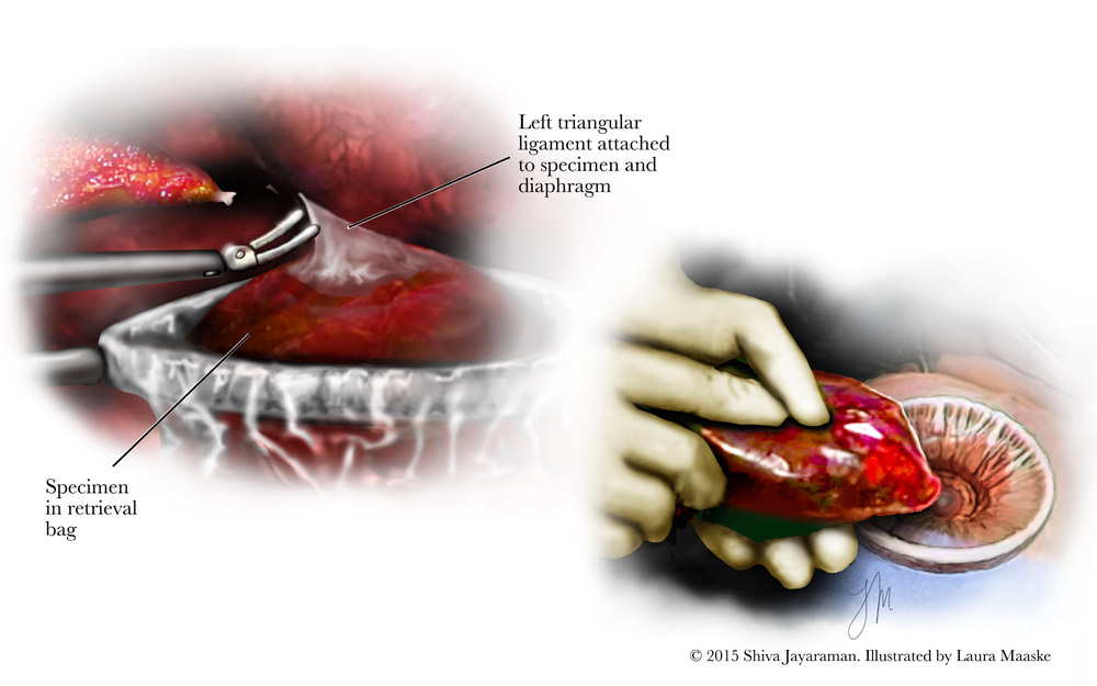
Some final attachments remain. And the left triangular ligament is still intact. This helps suspend the specimen making it easy to place the liver which must be removed into an endcatch bag. Bagging the specimen aids in the preventive spread of the liver tumor. When the specimen is in the bag a final cut is placed to release the liver segment from its attachment within the abdomen. The liver is extruded through a Pfannenstiel incision, which is a horizontal cut placed in the lower abdomen. Note the placement of this incision as illustrated on the first positioning slide above.
A video of Dr. Jayaraman’s Laparoscopic Left Lateral Liver Sectionectomy revealing the various steps and the surgical scene as seen by the surgeon:
Review:
Summary
This surgical medical illustration series reveals the Laparoscopic Left Lateral Liver Sectionectomy procedure, which is performed to remove a tumor and Dr. Jayaraman is seeking to improve a technique, guided by his personal experience as a surgeon, to enhance patient care. I’ve illustrated these liver surgery illustrations as a guide for professional audiences.
Suggested Tweet:
“An Illustrated Laparoscopic Left Lateral Liver Sectionectomy“
Laura Maaske, MSc.BMC.
Biomedical Communicator
Medical Illustrator
Medical Legal Illustrator
Medical Animator
Health App Developer


