Medical Slide: Isolation of Splenic Artery & Vein in Left Laparoscopic Distal Pancreatectomy. A retractor is lifting the pancreas away from the splenic artery and vein so these vessels can be preserved. Medical Illustration. Copyrighted Material Illustrated by Laura Maaske Medical Illustrator, Medical Animator, Biomedical Communicator, & biological artist. For permission information contact the artist

Illustrating the Lateral Approach to the Left Laparoscopic Distal Pancreatectomy

The Laparoscopic Distal Pancreatectomy is a surgical procedure performed to remove both solid and cystic tumors from the

Pancreas Medical Illustration Series: Laparoscopic Illustrations

Written and illustrated by Laura Maaske, MSc.BMC, Medical Illustrator & Medical Animator

These surgical illustrations are a reflection of the Lateral Approach to the Left Laparoscopic Distal Pancreatectomy as it is performed by Dr. Shiva Jayaraman, MD, MESc, FRCSC, of St. Joseph’s Healthcare Center, University of Toronto, Canada.


Overview of the Left Lateral Laparoscopic Distal Pancreatectomy

The Laparoscopic Distal Pancreatectomy is a surgical procedure performed to remove both solid and cystic tumors from the distal pancreas. Tumors of this type to this area of the pancreas usually affect middle-aged adult women. Surgeons aim to preserve as many of the critical splenic and pancreatic vessels as possible. Careful effort is made to preserve the surrounding tissues. This way, the blood supply to vital pancreatic tissues is not disturbed. The procedure is performed laparoscopically as this is less invasive than an open surgical technique, and offers a more cosmetic impact.

The greatest challenge in performing this surgery safely is that it is difficult to remove a tumor from the pancreas without the risk of damaging the spleen. While it is sometimes necessary to preserve the spleen without the splenic vessels being cut, whenever it is possible these vessels are kept intact. If the splenic vessels are cut, the surgeon must determine what to do with the remnant of the spleen to preserve its blood supply.


History of this Procedure

In the year 1911 Dr. Bernheim, at John Hopkins University, was the first surgeon to perform laparoscopy to explore a pancreatic tumor  But because the procedure is risky and splenic funtion was difficult to preserve, it wasn’t until 1994 that Dr. Soper removed a distal pancreatic tumor in a pig. Then in 1996 Dr. Gagner performed 5 laparascopic distal pancreatectomies on humans.
Patients are diagnosed based on ultrasound images, computerized tomography, an abdominal MRI, and a cholangiogram of Wirsung’s duct  The procedure below is performed by Dr. Shiva Jayaraman, at Universiry of Toronto. And you will see in this procedure that the splenic vessels are preserved.


Steps in the Procedure

I illustrated this series for Shiva Jayaraman of St. Joseph’s Health Center in Toronto, Canada. I created the illustrations particularly to follow his technique. For more information about Dr. Jayaraman’s approach, please see his article on the topic, Laparoscopic Distal Pancreatectomy: Evolution of a Technique at a Single Institution.

1. Patient Positioning

Patient Positioning. Left Laparoscopic Distal Pancreatectomy

Patient Positioning for Left Laparoscopic Distal Pancreatectomy


2. Mobilization of splenic flexure

(lesser omentum still intact. Pancreas visible behind omentum. Arrows on drawing reveal the act of mobilization of the lesser omentum)


Mobilization of splenic flexure in Left Laparoscopic Distal Pancreatectomy

Mobilization of splenic flexure for Left Laparoscopic Distal Pancreatectomy


3. The Distal Tip of the Pancreas is Exposed

Through a laparoscopic surgical approach the surgeon gains infragastric access. Using an atraumatic grasper introduced through the left paraxiphoid trocar, the stomach is lifted at the great curvature and raised using a grasper. The spleno-colic ligament is divided. The inferior border of pancreas is dissected away to reveal deeper anatomy. And the colon retracted away from the area of action by gravity.

Division of Spleno-colic Ligament &  Dissection of Inferior Border of Pancreas in Left Laparoscopic Distal Pancreatectomy

 Division of Spleno-colic Ligament & Dissection of Inferior Border of Pancreas  for Left Laparoscopic Distal Pancreatectomy


4. Isolation of splenic artery and vein while peeling pancreas

Next, the splenic vein is isolated in order to allow its preservation. As the pancreas itself is attached to the vein and artery, it is carefully dissected away in a lateral to medial direction using hook cautery.
Isolation of Splenic Artery & Vein in Left Laparoscopic Distal Pancreatectomy

Isolation of Splenic Artery & Vein in Left Laparoscopic Distal Pancreatectomy

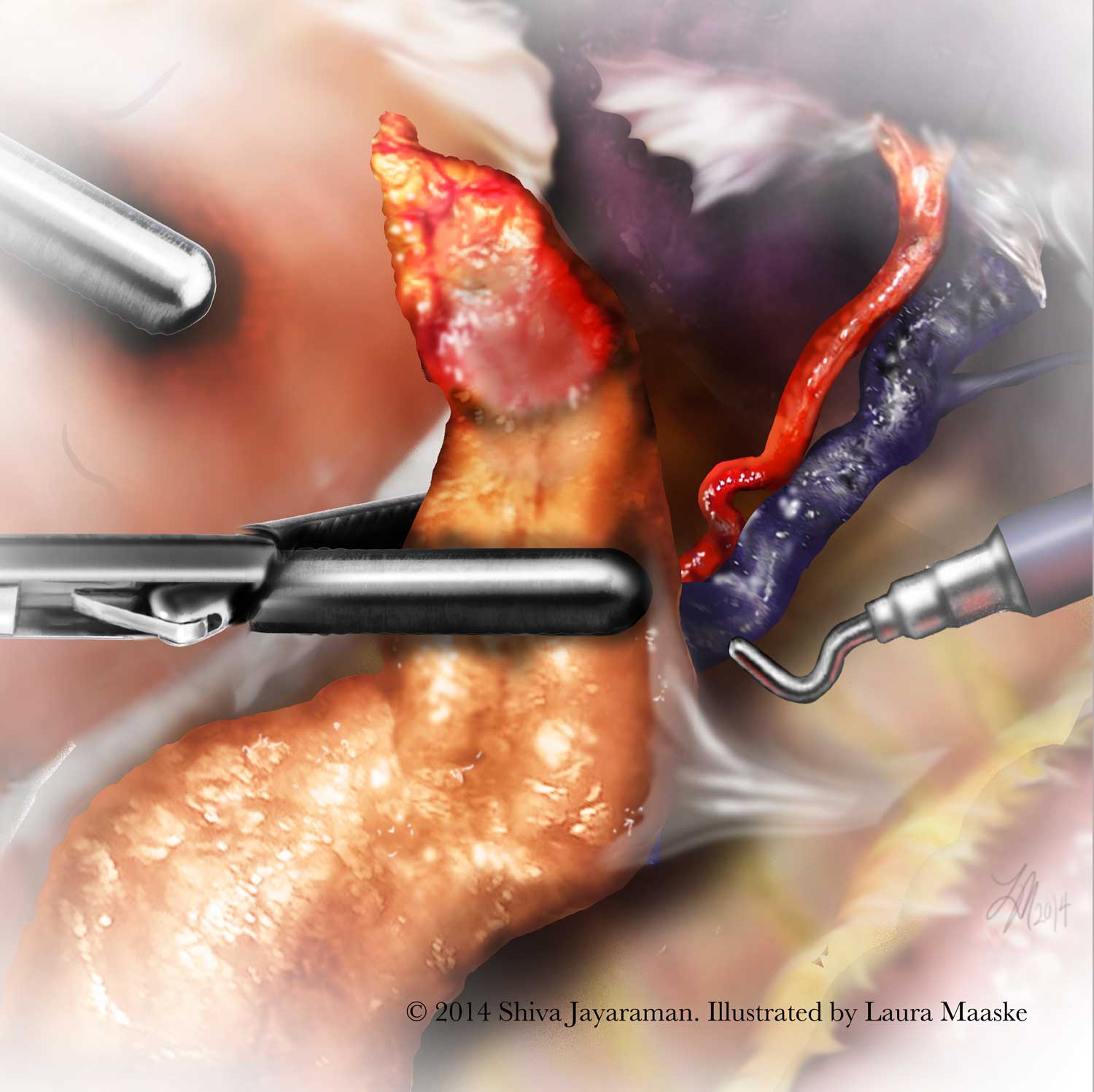
 Isolation of Splenic Artery & Vein for Left Laparoscopic Distal Pancreatectomy


5. Post Pancreatic Resection

The pancreas is then divided to isolate the tumor. Note below the pancreatic stump with a staple line in view. Note also that the vessels are intact as a result of careful dissection.
About the illustrations: Generally I begin my surgical drawings with rough pencil sketches for the ideas to be covered in the procedure. Anatomy for laparoscopic work is tight, but nonetheless very complex. Although I have  strong background in anatomy, each region of the body presents a complicated three-dimensional anatomical field. So I review the details by referring to various texts. I love watercolors and sometimes I begin a surgical painting there. But in this case, I rendered the sketches entirely using Adobe Photoshop. Final pieces are used digitally in this case. Alternatively, I may print the pieces on watercolor paper with nice results. Please feel free to contact me with any questions.

Resection of Pancreas to Remove Tumor in Left Laparoscopic Distal Pancreatectomy

Resection of Pancreas to Remove Tumor for Left Laparoscopic Distal Pancreatectomy

This step completes the Left Laparoscopic Distal Pancreatectomy procedure.


For more information, see Dr. Jayaraman Youtube Channel. All my thanks to Dr. Jayaraman for his patience in offering details of his laparoscopic procedure, and for his succinct surgical explanations.


Please try my preworded tweet to share this article:

An Illustrated Novel Approach to the Distal Pancreatectomy with Shiva Jayaraman 


January 30, 2015

Laura Maaske, MSc.BMC. Biomedical Communicator

medimagery_medical-illustrator-350
????????????????????????????
Laura Maaske – Medimagery LLC
Medical Illustration & Design
????????????????????????????
Pin It

About Laura

Medical Illustrator; science and art lover.