
Male Pelvic Anatomy
The male pelvis depends on these tissues to achieve its functions of reproduction and urinary excretion:
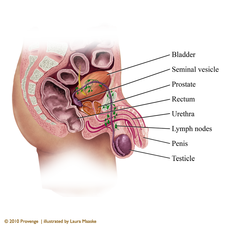
- bladder - a muscular and layered sac resting just above and posterior to the pubic bone. When empty, the bladder is about the size and shape of a pear. As the kidneys manufacture urine, the fluid passes through the two ureters and empties into the bladder. The bladder stores urine until it can be voluntarily excreted via two sphincters (valves), which release the urine into the urethra. The bladder muscles contract and urine flows through the urethra. During urination, the bladder muscles contract, and open to allow urine to flow out. Urine exits the bladder into the urethra, which carries urine out of the body.
- urethra - a duct that runs through the center of the prostate, from the bladder through the penis, and allows urine to exit the body. The urethra is much longer in males than in females, because of its passage through the penis. It is 8 inches long in males and 1.5 inches long in females.
- penis - required for urinary excretion and sexual copulation. Erection occurs when erectile tissue in the penis swells and becomes engorged. The arteries dilate. But the veins collapse so that blood is held within the tissue.
- scrotum - a pouch hanging behind the penis to hold and protect the testes. The testes depend on a specific temperature range to function optimally, to which the many nerves and blood vessels in the scrotum are sensitive. When the temperature is too low, the cremaster muscle contracts and pulls the testes closer to the body. The inguinal canal connects the scrotum to the abdomen. Within the inguinal canal lie the spermatic cord and its connective tissue which holds the spermatic artery, vein, nerve.
- epididymis - this tissue, cupped around the top of the each testis, holds the sperm before they pass to the vas deferens. Many tubes within the epididymis hold the sperm while they mature and move on to the ampullary gland and prostatic ducts.
- vas deferens - is a thin, tubular, sperm duct. The vasa deferentia (singular: vas deferens) bring sperm from the testes to the seminal vesicles. The seminal vesicles contribute fluid to semen during ejaculation.
- There are 3 accessory glands which provide fluids to lubricate the ducts and nourish the spermatazoa:
- prostate - is a gland that surrounds the ejaculatory ducts at the base of the urethra before it enters the penis. The prostate is located behind the pubic bone, above the perineal membrane and penis, below the bladder, and in front of the rectum. The prostate produces a slightly basic fluid, semen, by mixing calcium with semen coming from seminal vesicle in a process called profibrinolysin. The fluid nourishes and protects sperm. During the first stages of ejaculation, the smooth muscles of the prostate squeezes this fluid into the urethra where it mixes with semen. Sperm that are expelled with semen from the prostate are far better at moving to their destination than sperm mixed only with seminal vesicular fluid. As men age, the size of their prostates changes.
- seminal vesicles - are pouches attached to the vas deferens at one side of the bladder. They produce a sticky, yellowish fluid that contains energy sperm need to for their movement. Most of the semen is made up of this fluid.
- bulbourethral glands (Cowper glands) - are two small glands that lie along the urethra just below the prostate gland, with ducts connecting to the urethra. They produce a clear, shiny fluid.
Laura Maaske, B.Sc., M.Sc.BMC
Racine, WI 53402
Cell: (262) 308-1300
Contact Laura
Text Copyright © Medimagery - Laura Maaske LLC
Illustration © 2010 Provenge, illustrated by Laura Maaske - Medimagery LLC.
See also the Male Pelvic Artery Illustration here on this site.
Return to Male Pelvis Anatomical Illustration.


We specialize in highly interactive dynamic medical illustrations for both print and e-publishing. Content is dynamic and interactive, or traditional. Illustrations are prepared for advertising, pharmaceutical, publishing, health promotion, health professional education, children, and medical-legal resources. All medical, e-book illustrations, dynamic and conceptual artwork are prepared by hand for the client's unique needs. All materials Copyright © - Laura Maaske - Medimagery LLC. Call Laura at 262-308-1300 with questions or to request a price quote.