


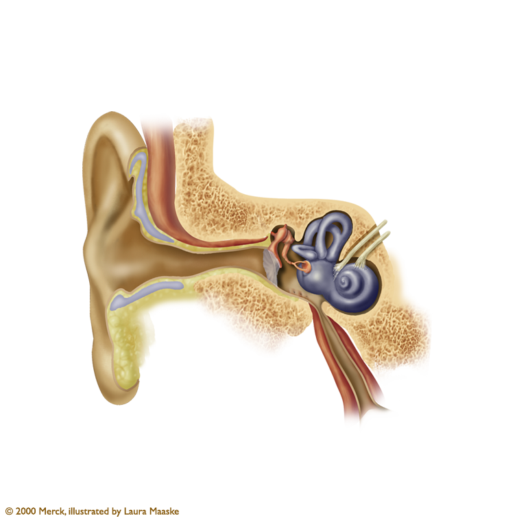
Ear Anatomy
The pinna (or auricle) is the part of the ear we can see, the outer cartilage. Sound enters the ear and is directed through outer ear canal. Sound vibrates the ear drum (tympanic membrane), which vibrates the three bones of the Middle ear: from hammer (malleus) to anvil (incus) to stirrup (stapes). The stapes is, incidentally, the smallest bone in the human body. This vibration is passed to the Inner Ear, to a spiraled structure called the cochlea. The lining of the cochlea, a structure shaped like a snail and filled with fluid, is comprised of tiny cilia. The movement of these cilia send neural impulses to various parts of the brain for the interpretation of sound.
Another structure, the eustachian tube, is important for equalizing pressure between the Middle Ear and the outside air. It is a tube connecting the middle ear to the back of the nose.
There are three fluid filled, looped structures in the Inner Ear. They are attached to the cochlea and, based on the turn of the head, they send signals about balance and dizziness.
© 2000 Merck Pharmaceutical, illustrated by Laura Maaske - Medimagery LLC
(click the plus in the upper right corner to return to the illustration)


We specialize in highly interactive dynamic medical illustrations for both print and e-publishing. Content is dynamic and interactive, or traditional. Illustrations are prepared for advertising, pharmaceutical, publishing, health promotion, health professional education, children, and medical-legal resources. All medical, e-book illustrations, dynamic and conceptual artwork are prepared by hand for the client's unique needs. All materials Copyright © - Laura Maaske - Medimagery LLC. Call Laura at 262-308-1300 with questions or to request a price quote.