
Medical Illustration of the Eye: Cataracts and their History through Art
Human Vision: Cataract of the Eye
The word “cataract” has Latin and Greek origins, meaning “waterfall” and “downpour”. Cataracts were thought in those times to have arisen from an outpouring of corrupt humour ino the eye. The word may have been used to metaphorically describe the appearance of white clouds in the eye, the same as churning water.
In the United States, half of Americans over age 65 have a cataract, According to the Word Health Organization (WHO). And among the citizens of the world at large, cataracts are the number one cause of blindness.
What are Cataracts?
Cataracts are generally defined as breakdown of the lens microarchitecture. Normally in vision, light passes through the lens of the eye so that the image can be projected clearly on the retina. This image is then interpreted as a neural signal. If the lens is unclear, which is a condition called a cataract, the image projected on the retina will be cloudy or blurred. With a cataract, some even, perhaps a DNA mutation, causes opacity of the crystalline lens. It may be there isa structure change accompanied by changs inin lens protein constituents. The opacity or cloudiness means that the refractive index of the lens varies significantly over distances approximating the wavelength of the transmitted light.
Aside from blurriness, symptoms of cataracts may include one or many of these:
- Light sensitivity
- Glare or hazy vision
- Night vision loss
- Halos appearing around light sources
- Flashes of light
- Double vision in one eye or both
- Seeking brighter light for tasks which previously did not require it
- Objects in the visual scene appear less vivid, yellowish, or dull
- Several changes in prescription strengths
- Poor sleep
Cataracts are common and occur naturally with aging and usually develop slowly, almost imperceptibly. But they may also develop as a result of injury. Some infants are born with them. Other causes of cataracts include excessive exposure to sunlight, diabetes, hypertension, smoking, obesity, radiation and ultraviolet light exposure, previous eyesurgery, obesity, excessive alcohol use, inadequate Vitamin C, or longtime usage of corticosteroid medicines. Historically, arsenic poisoning caused cataracts and may have even been the source of Jane Austen’s poor vision. There is a genetic origin to cataracts and it is believed the cataract begins with a DNA mutation.

Types of Cataracts
Cataracts can be classified by a LOCS III system in which cataracts are categorized by severity on a scale of 1 to 5, and by on of three types:
- Nuclear sclerosis is common. A person with this type of cataract may experience increasing near sightedness. The cataracts begins as a cloudy formation in the central, nuclear region of the lens. Later the formation becomes hard (sclerotic) As the nuclear sclerosis develops it is called a brunescent cataract.
- Cortical cataracts happen when the outer layer of the lens, the lens cortex, becoming opaque. People with this type of cataract are especially susceptible to glare and night vision compromises. Liquid at the periphery of the lens causes fissuring. When these cataracts are viewed through an ophthalmoscope, or magnifier, there is a characteristic appearance like the spokes of a wheel.
- Posterior subcapsular cataracts demonstrate a cloudy deposit at the back of the lens. As a result of lens magnification in the eye, the cataract appears larger and is more obstructive to the sufferer. And symptons, as well, are magnified.
All cataracts with cloudy deposits begin when the cloudy proteins are more transparent and become more obstructive as the proteins mature.
What is the Treatment for Cataracts?
If a cataract is interfering with vision significantly, it can be removed in a surgical procedure.
Standard Surgical Procedure
The eventual treatment for cataracts, when they begin to interfere with everyday tasks, is typically surgery. An ophthalmologist will use a surgical procedure, with the assistance of ultrasound, to make a tiny cut in the eye to remove the lens and replace it with a clear plastic lens. The artificial lens is called an intraocular lens (IOL).
Immediate complications of surgery may include:
- Endophthalmitis, or infection in the eye
- Retinal detachment, or detachment of the back retina of the eye
- Cystoid macular edema, which is swelling and fluid in the center of the nerve layer
- Corneal edema, which is swelling of the clear covering of the eye
- Hyphema, or bleeding of the eye
Long term complications may include:
- Retinal detachment
- Posterior capsular opacification, or aftercataract. This is a cloudiness in the vision which remains after surgery and is often treated by laser.
- Strabismus, or an abnormal alignment in the eyes
- Glaucoma, or increased pressure within the eyeball
- Ptosis, or a saggy upper eyelid
- Glare in vision
- Dislocated intraocular lens
- Astigmatism, or a defect in spherical curvature of the lens itself, resulting in distorted images in the vision
History
Historical Treatments of Cataracts
Treatment for cataracts traces back almost 4000 years.
2467 B.C.E.
Cataracts are such a common human experience, it is not surprising that there are plenty of references in historical documents. While it is presumed cataract treatments are ancient beyond recorded history, the earliest artistic depiction of a cataract was created in Egypt around 2457-2467 B.C.E. It is a small wooden statue of an Egyptian priest treating a patient with a white pupil, representing the cloudy lens.
1754 B.C.E.
The earliest documentation of cataract treatment dates to Egypt. In the Code of Hammurabi, a Babylonian law code of ancient Mesopotamia created in 1754 B.C.E., the law dotes out payment and the punishment for surgeons if the patient were to die or lose their vision. The surgeon’s finger is to be cut off (from, The Eye in History by Frank Joseph Goes, p. 371)
500 B.C.E | Couching Procedures for Cataracts
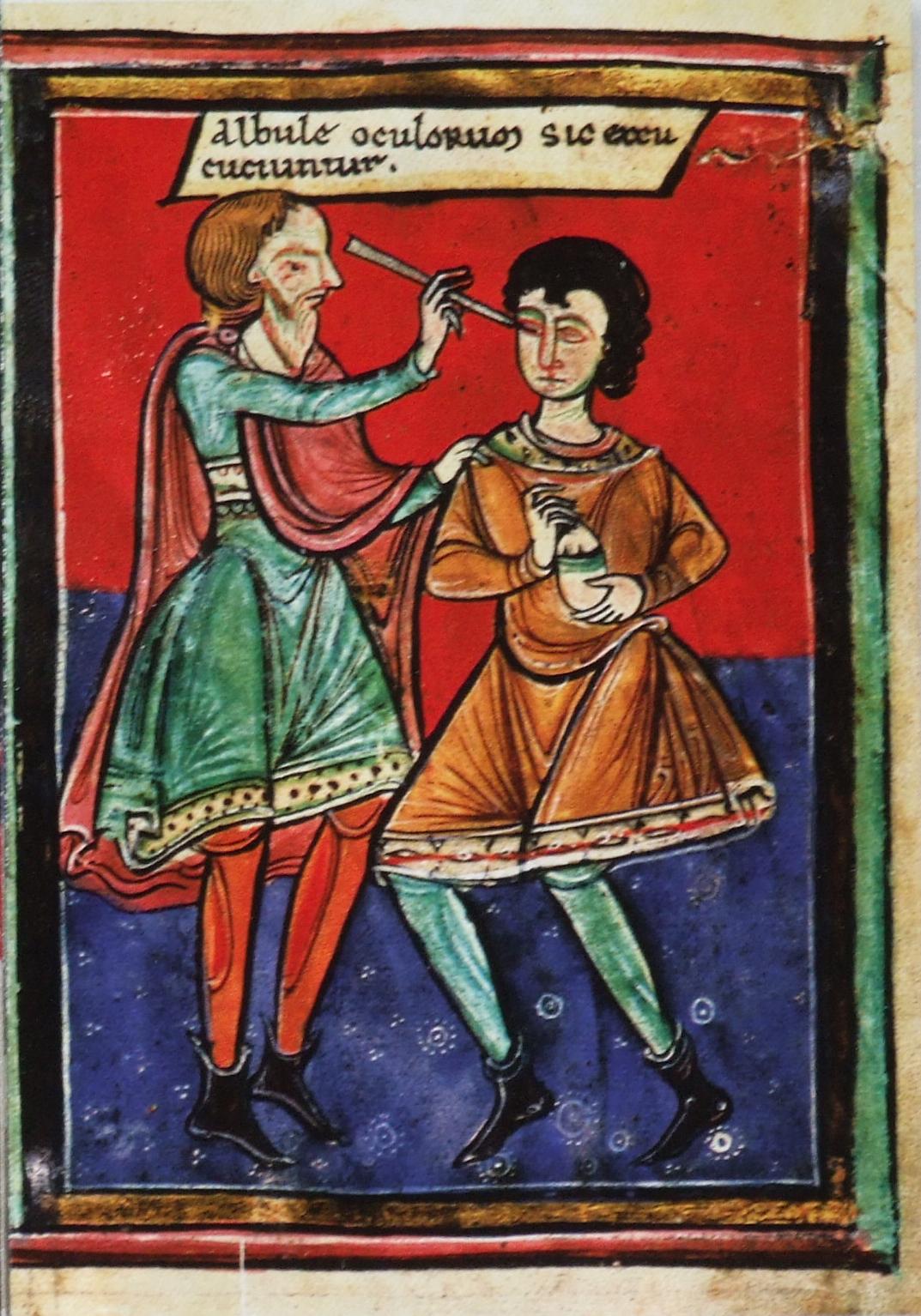
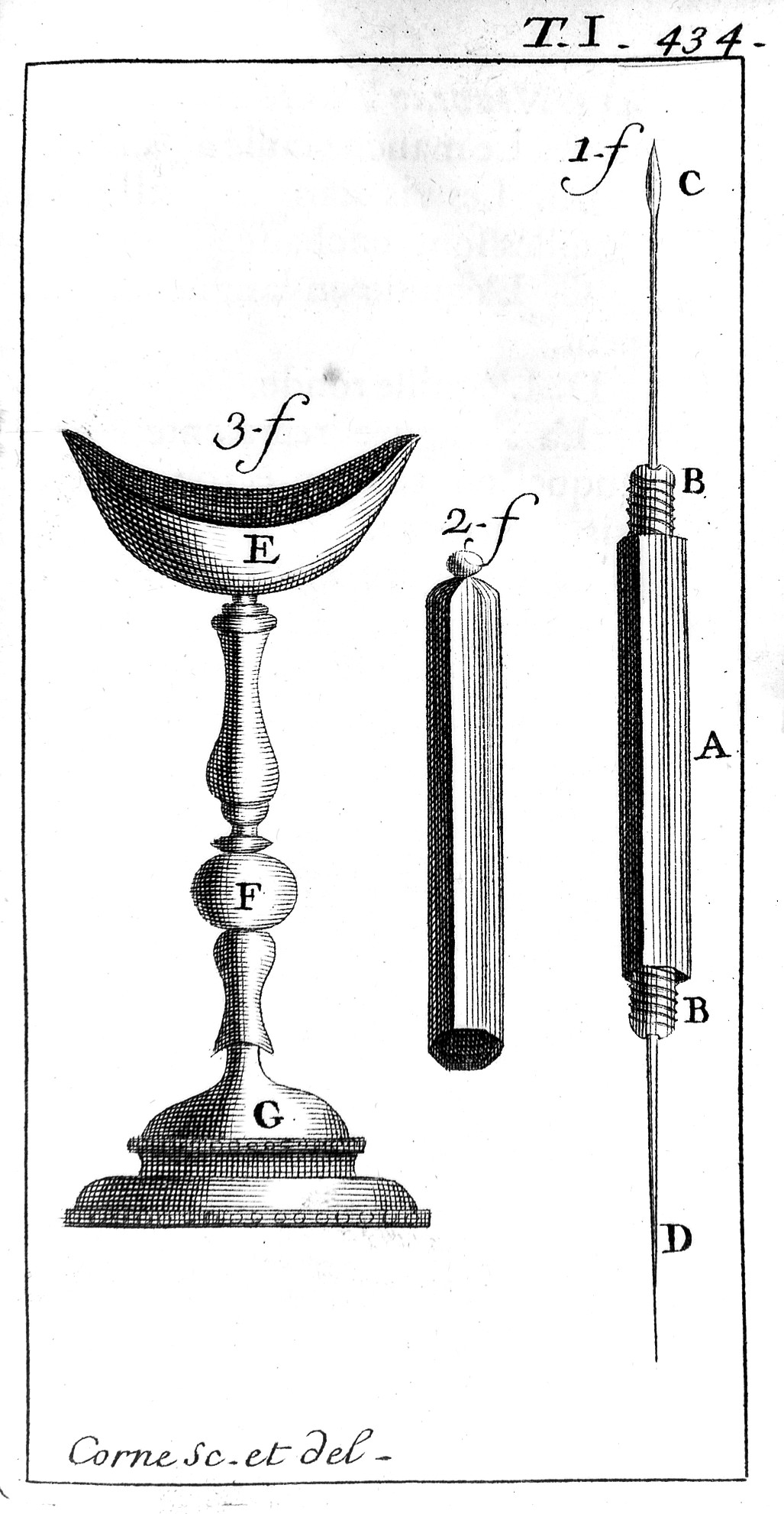
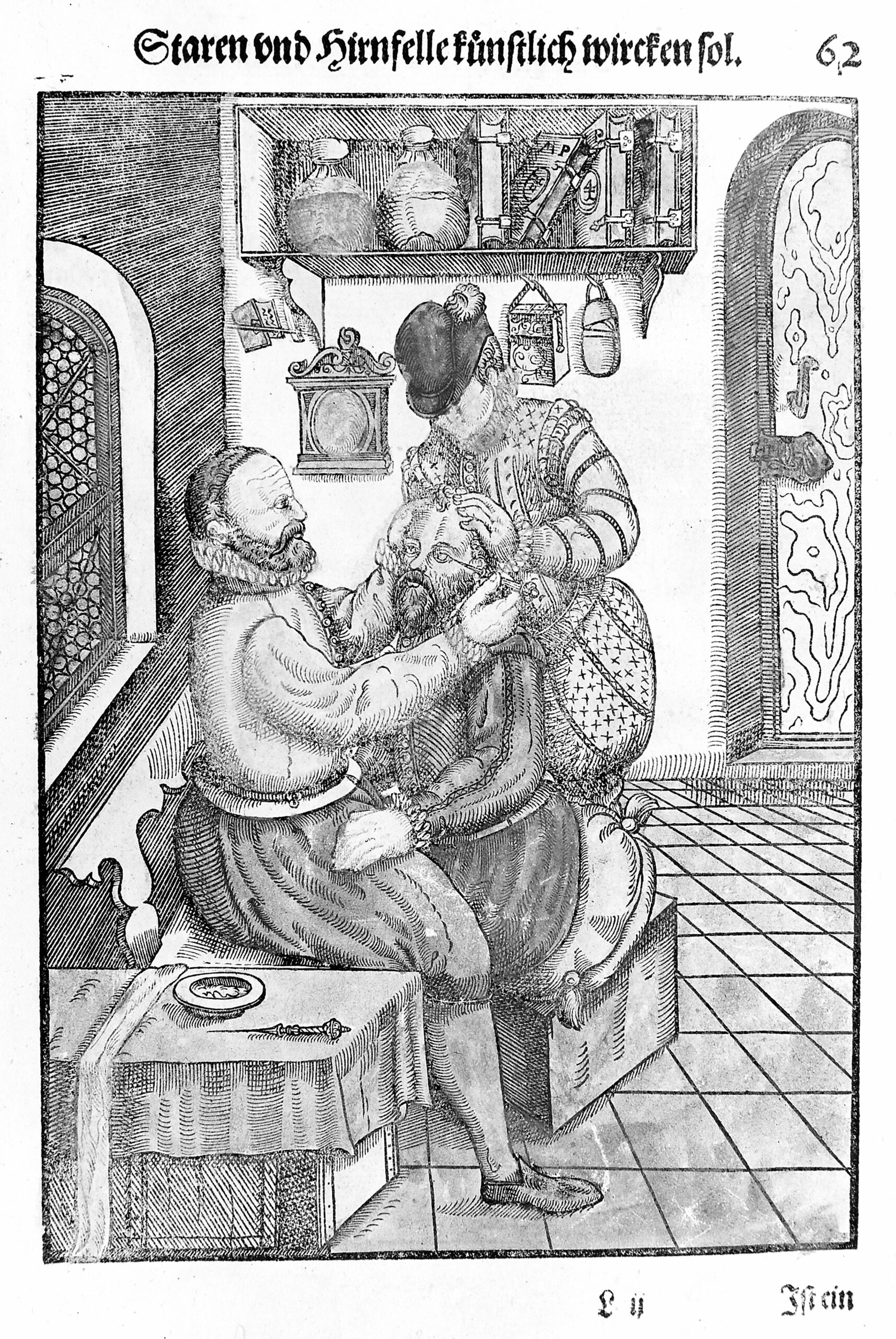
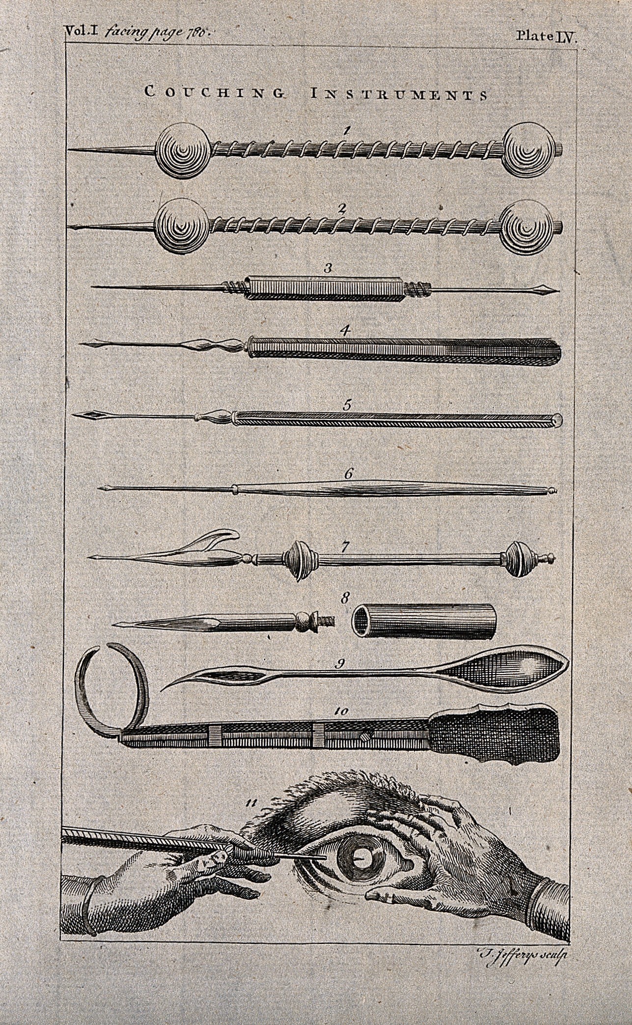
The Chinese, the Indians, the Egyptians, the Romans, and the Greeks all carried out surgical procedures on cataracts. The treatment used through most of human history is known as couching. There are two main types of couching:
- Dating to around 500 B.C.E. the procedure was only useful for those who were blinded by severe cataracts, in which the lens had become rigid and entirely opaque, where the lens capsule and zonules have weakened their hold of the lens. This involved hitting the patient’s eye with a blunt object so the lens fell backward into the vitreous humor in the back of the eye. The lens would eventually be reabsorbed into the body and the patient’s eye would allow in light, but visual would be extremely blurry.
- By 29 A.D. Western physicians had begun to use the sharp end of a needle was inserted into the eye to poke lens out of its placement a the iris and into the eyeball itself. And as time passed, surgeons learned to break the lens into pieces so it would more easily be absorbed by the vitreous humor. Again, the procedure resulted in blurry vision with greater capacity to let in light. The blunt end of the needle was used to cauterize the incision when the needle was used.
There were no anaesthetics. An assistant held the patient still. Couching is still performed in some places around the world today.
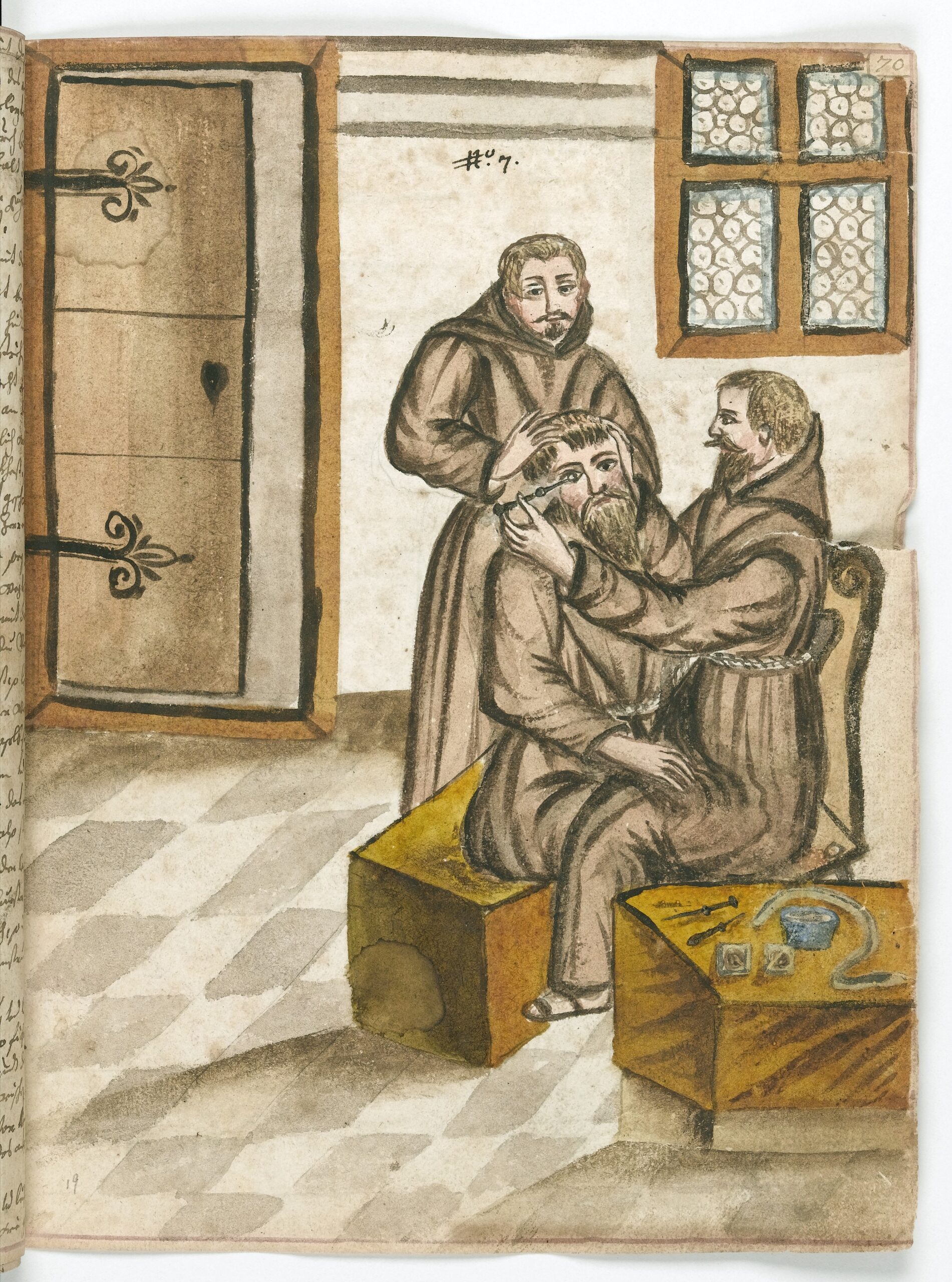
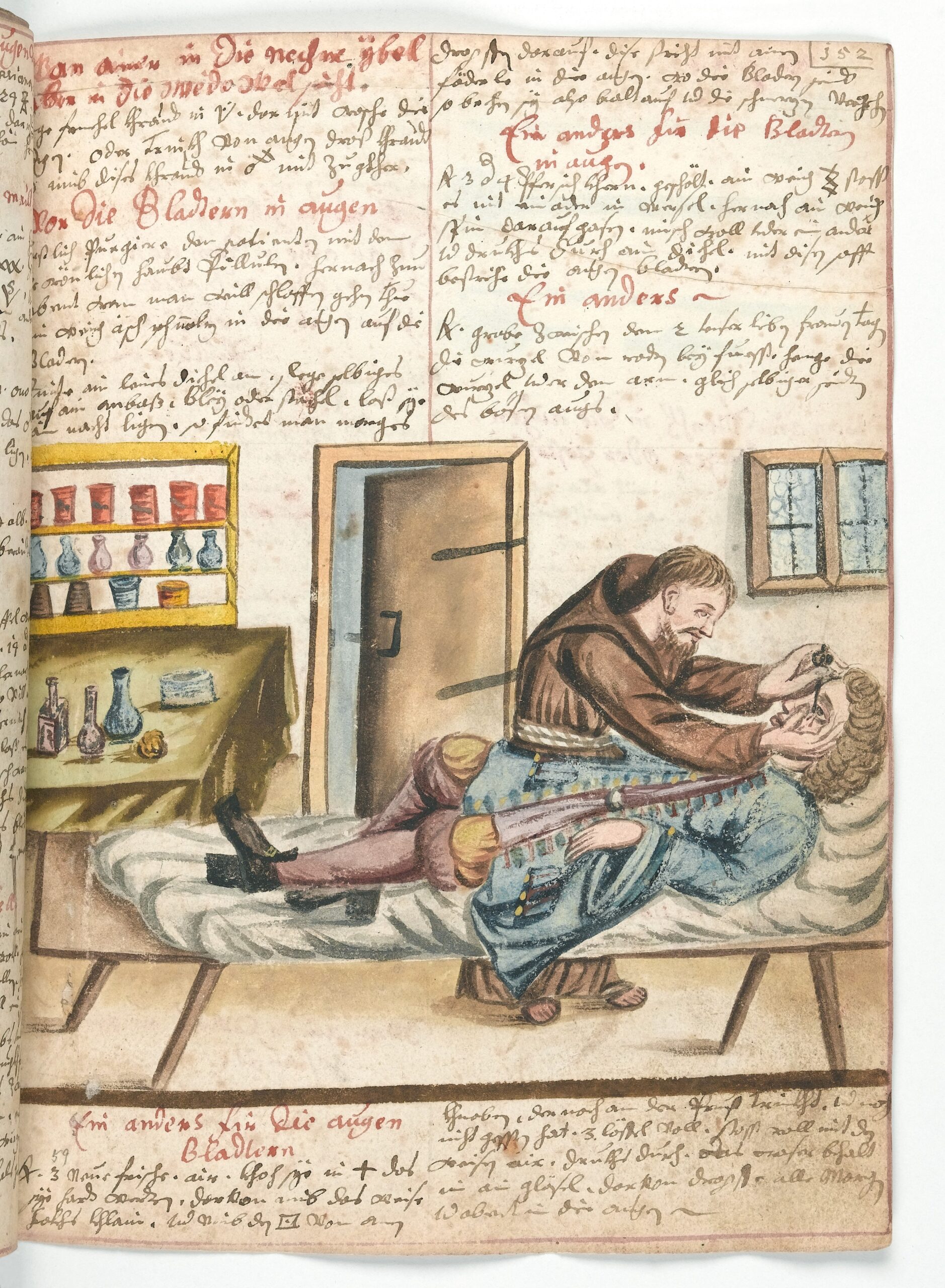
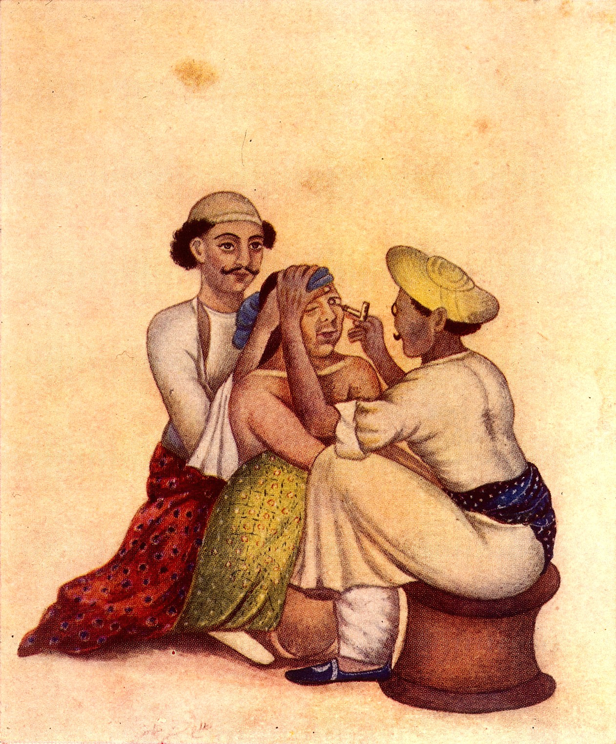
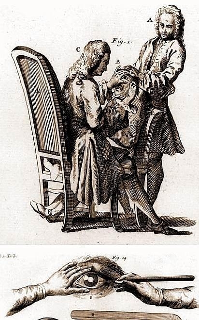
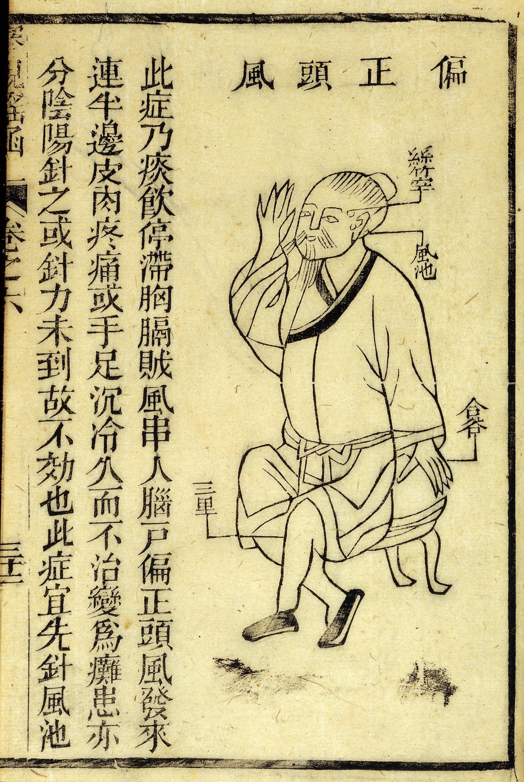
1753 A.D. | Extracting the Lens rather than to use Couching
Parisian Jacques Daviel was the first physician to remove the lens for cataract procedures, in 1748. He made an incision in the eye to release the lens outwardly. Samuel Sharpe of London, England used a specific thumb technique, applying pressure to pop out the lens, in 1753. By 1902 physicians had begun using suction cups and forceps to extract the lens.
1761 A.D. | Cataract Procedures in the Colonial Period
Early settlers in North America presumably had greater survival concerns than cataracts. Those who could afford the surgery returned to England or other European countries for the surgery. Or to the Caribbean after 1751.
Dr. Stork is the first known surgeon to practice cataract removal procedures in the United States. He advertised his skill in, ?the Art of restoring to Sight, and curing the different Diseases of the Eye.” in Philadelphia, in 1761.
Unfortunately, anaesthesia is still not in use at this time and was not until 1840.
1949 A.D. | Discovery of the Artificial Lens
Early in the treatment of contacts, ophthalmologist experienced with glass lenses as lens replacements. But these were rejected by the eye as foreign matter. During World War Two pilots were injured frequently by the shattering of acrylic cockpit glass. The shards of acrylic would occasionally lodge in the eyes of pilots. In 1946 Sir Harold Ridley was the ophthalmologist who noticed that the eye did not reject acrylic in the eye, and proposed the treatment of cataracts with artificial lenses. Cataract surgery quickly became a meaningful surgical treatment for cataracts. In 1949 the first artifical lens procedure was performed, and was a success.
1957 A.D. | Alpha-chymotrypsin for Removal of Lens
Jose Barraquer of Spain used alpha-chymotrypsin to enzymatically dissolve the zonules for removal of the lens, in the year 1957. He also developed an ophthalmological instrument, the Barraquer Keratotome with pneumatic fixation, to perform more precise cataract incisions. He used an air injection technique in the anterior chamber in cataract surgery. In fact, Jose’s father was Ignacio Barraquer, who is also famous for having made innovations in cataract surgery including the development of the suction cup for removing the pieces of the lens.
1961 A.D. | Cryo-surgery for Removal of Lens
Tadeusz Krwawicz of Poland invented a cryosurgical technique attaches a tiny probe, a silver rod conducting cold from solid CO2 in a tiny syringe, to the eye by freezing a small area on the surface of the lens to extract the lens. The probe was called a cryoextractor, and allowed adhesion to the lens to be more reliable.
1967 A.D. | Phacoemulsification Technique
In 1967, New Yorker Charles Kelman developed a far less invasive and less painful technique using ultrasonic vibrations to break up the lens of the eye into tiny particles which are subsequently sucked from the eye through the incision.
1986 A.D. | Laserphaco Probe
Patricia Bath (the first African American to complete a residency in ophthalmology, in 1973) invented the Laserphaco Probe in 1986 to improve the accuracy in cataract procedures of breaking down the lens through laser treatment and a 1mm incision. The 1986 technique is called, phacoblation.
2016 A.D. | Stem Cells for Human Lens Regeneration in Cataracts
In 2016 the Journal Nature published a study using an animal’s own stem cells to treat cataracts. The researchers successfully removed cataracts in infants (and rabbits) while preserving epithelial stem/progenitor cells (LECs). These stem cells have an important role in lens regeneration. In these cases, the lenses regenerated from the stem cells.
They say this method not only allows the lens to regenerate naturally but is less surgically invasive than current treatment methods. According to the research, recovery and vision in these infants, rabbits, and macaques, was superior to patients who received the standard surgical procedure for cataracts and the artificial lens. This stem cell is expected to be effective in other age groups as well, but no new research on this subject has been released, to date.
Famous People with Cataracts
The great artist Claude Monet underwent two surgeries for his cataracts. Both occurred in 1923, when he was 83 years old. One scientist analyzed the deterioration in Monet’s vision through his paintings here.
Jane Austen may have suffered from cataracts, as well; revealed in descriptions of her vision problems in letters written to friends.
Perspective
Humans have a long history of treating and working with the fairly common experience of cataracts. Recent treatments are far more successful, The newest stem cell treatment, on the horizon for the majority of sufferers, creates a feeling of great expectation in the long-time human endeavor for treatment.
May 3, 2017
Laura Maaske, MSc.BMC. Biomedical Communicator

Illustration Copyright © 2000 Merck Pharmaceutical, illustrated by Laura Maaske – Medimagery LLC
Text Copyright © 2017 Medimagery – Laura Maaske LLC
[jp_post_view]


I love doing the research and I’d be happy to add more. Are there any subtopics you are particularly seeking?
Laura